Review
. 2017 Oct;15(6):522-534.
doi: 10.2450/2017.0353-16. Epub 2017 May 15.

Influence of red blood cell-derived microparticles upon vasoregulation

Affiliations
Review

Influence of red blood cell-derived microparticles upon vasoregulation

Ahmed S Said et al. Blood Transfus. 2017 Oct.

Abstract

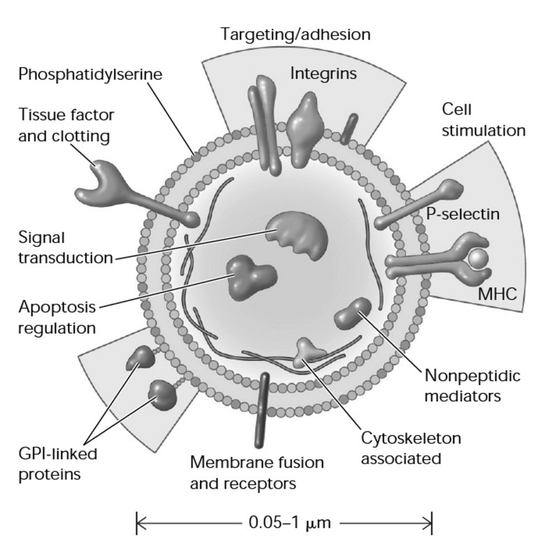
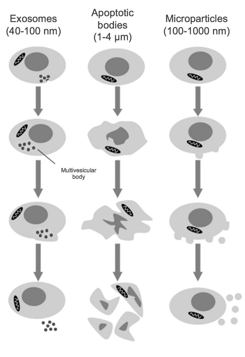
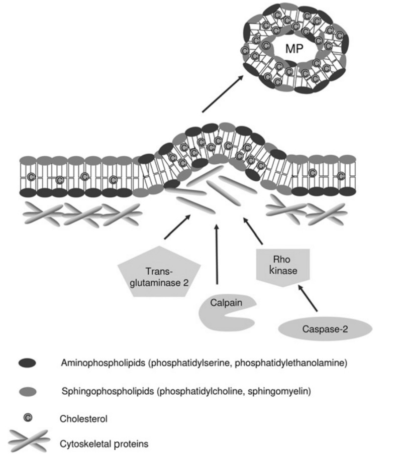
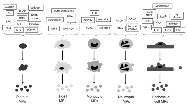
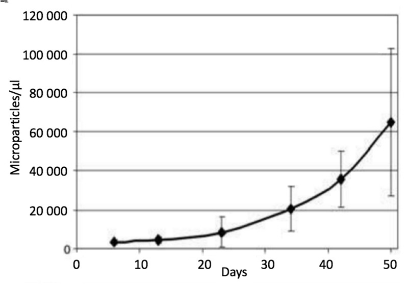
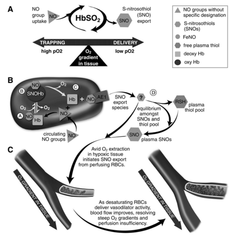
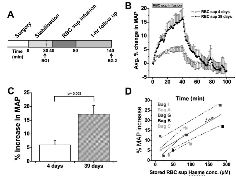
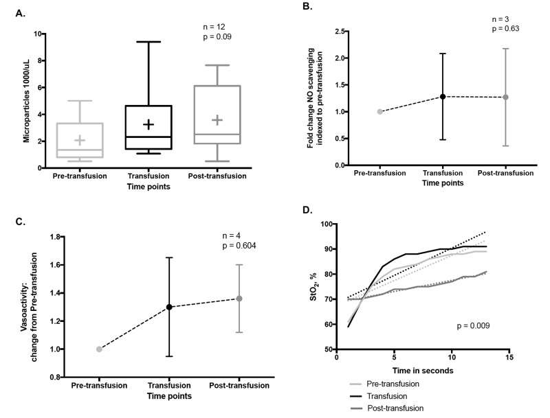
Here we review recent data and the evolving understanding of the role of red blood cell-derived microparticles (RMPs) in normal physiology and in disease progression. Microparticles (MPs) are small membrane vesicles derived from various parent cell types. MPs are produced in response to a variety of stimuli through several cytoskeletal and membrane phospholipid changes. MPs have been investigated as potential biomarkers for multiple disease processes and are thought to have biological effects, most notably in: promotion of coagulation, production and handling of reactive oxygen species, immune modulation, angiogenesis, and in apoptosis. Specifically, RMPs are produced normally during RBC maturation and their production is accelerated during processing and storage for transfusion. Several factors during RBC storage are known to trigger RMP production, including: increased intracellular calcium, increased potassium leakage, and energy failure with ATP depletion. Of note, RMP composition differs from that of intact RBCs, and the nature and composition of RMP components are affected by both storage duration and the character of storage solutions. Recognised RMP bioactivities include: promotion of coagulation, immune modulation, and promotion of endothelial adhesion, as well as influence upon vasoregulation via nitric oxide (NO) scavenging. Of particular relevance, RMPs are more avid NO scavengers than intact RBCs and this feature has been proposed as a mechanism for the impaired oxygen delivery homeostasis that has been observed following transfusion. Preliminary human studies demonstrate that circulating RMP abundance increases with RBC transfusion and is associated with altered plasma vasoactivity and abnormal vasoregulation. In summary, RMPs are submicron particles released from stored RBCs, with demonstrated vasoactive properties that appear to disturb oxygen delivery homeostasis. The clinical impact of RMPs in transfusion recipients is an area of continued investigation.


Conflict of interest statement

The Authors declare no conflicts of interest.

Figures

Figure 1
Figure 1
Cellular microparticles: a mobile storage pool of bioactive effectors. Membrane microparticles are shed from the plasma membrane of stimulated cells, harbouring cytoplasmic proteins as well as bioactive lipids implicated in a variety of fundamental processes. MHC: major histocompatibility complex; GPI: glycosylphosphatidylinositol. (Adapted with permission from Hugel et al., 2005.)
Figure 2
Figure 2
Size classes of extracellular vesicles. Exosomes are formed through inward membrane budding, leading to formation of 40–100 nm intracellular vesicles which accumulate within multivesicular bodies that are subsequently released to the extracellular milieu. Apoptotic bodies may contain DNA and/or organelles and are formed during the late stages of apoptosis, after cell shrinkage. Microparticles are formed from the outward blebbing of membrane and released into the extracellular space. (Adapted with permission from Burger et al., 20133).
Figure 3
Figure 3
Mechanisms proposed for cytoskeleton remodelling leading to microparticle (MP) formation. Under normal conditions, aminophospholipids (phosphatidylserine and phosphatidylethanolamine) are found exclusively on the inner leaflet of the plasma membrane. During MP formation, membrane asymmetry is lost as aminophospholipids redistribute to the outer leaflet of the plasma membrane. Cytoskeletal re-organisation results in the outward blebbing of the plasma membrane and may be dependent upon actin polymerisation, caspase 2/Rho kinase, calpain and/or transglutaminase 2. Such processes may vary between different cell types. MP formation appears to occur selectively in lipid-rich microdomains (lipid rafts/caveolae) within the plasma membrane. (Adapted with permission from Burger et al., 20133). MP: microparticles.
Figure 4
Figure 4
Stimuli for microparticle (MP) formation from platelets, endothelial cells and leucocytes. A summary of the stimuli which promote MP formation from platelets, endothelial cells and leucocytes. A23187: calcium ionophore A23187; TRAP: thrombin receptor activating peptide; NE: norepinephrine; ROS: reactive oxygen species; EPO: erythropoietin; IL-6: interleukin 6; TNFα: tumour necrosis factor α; LPS: lipopolysaccharide; sCD40I: soluble CD40 ligand; PMA: phorbol 12-myristate 13-acetate; FMLP: formyl-methionyl-leucyl-phenylalanine; ANCA: anti-neutrophil cytoplasmic antibodies; CRP: c-reactive protein; Ang II: angiotensin II; IL-1α: interleukin 1α; PAI-1: plasminogen activator inhibitor-1. (Adapted with permission from Burger et al., 20133).
Figure 5
Figure 5
Microparticle (MP) count in red blood cell units during storage (without centrifugation). Data are expressed as the mean±SD (n=7). At day 5: 3,371±1,188 MPs/μL were counted, whereas at day 50: MPs had approximately 20-fold (64,858±37,846 MPs/μL). MPs were stained with anti-human CD47. (Adapted with permission from Rubin et al.,71).
Figure 6
Figure 6
Red blood cells (RBC) transduce regional O2 gradients in tissue to control nitric oxide (NO) bioactivity in plasma by trapping or delivering NO groups as a function of haemoglobin (Hb) O2 saturation (Hb SO2). (A) Circulating NO groups are processed by Hb into the highly vasoactive (thiol-based) NO congener, S-nitrosothiol (SNO). By exporting SNOs as a function of Hb deoxygenation, RBCs precisely dispense vasodilator bioactivity in direct proportion to the lack of regional blood flow. (B) O2 delivery homeostasis requires biochemical coupling of vessel tone to environmental cues that match perfusion sufficiency to metabolic demand. Because oxygenated Hb (oxy Hb) and deoxygenated Hb (deoxy Hb) process NO differently, allosteric transitions in Hb conformation afford context-responsive (O2-coupled) control of NO bioavailability, thereby linking the sensor and effector arms of this system. Specifically, Hb conformation governs the equilibria among (A) deoxygenated Hb FeNO (NO sink), (B) oxygenated SNO-Hb (NO store), and (C) acceptor thiols including the membrane protein SNO-AE-1 (bioactive NO source). Direct SNO export from RBCs or S-transnitrosylation from RBCs to plasma thiols (D) yields vasoactive SNOs, which influence resistance vessel caliber and close this signalling loop. Thus, RBCs either trap (A) or export (D) NO groups to optimise blood flow. (C) NO processing in RBCs (A and B) couples vessel tone to tissue PO2; this system subserves hypoxic vasodilation in the arterial periphery and thereby calibrates blood flow to regional tissue hypoxia. (Adapted with permission from Doctor and Stamler, 2011. ©American Physiological Society). RSH: peptide or protein containing a thiol group.
Figure 7
Figure 7
Vasoactivity of infused packed red cell supernatant/plasma. (A) Experimental time line for packed red cell supernatant infusions. Rats were stabilised for 30 minutes (min) after surgery, and blood gasses were drawn as indicated (BG 1 and BG 2). Supernatant (1.6 mL) of packed red blood cells stored for either 4 or 39 days was infused for 40 min, after which the rats were followed for 1 hour (n=5). (B) Change in mean arterial pressure (MAP) over time after packed red blood cell (RBC) supernatant infusion and 60-min follow up. (C) Average percentage peak increase in MAP after infusion of packed RBC supernatants (RBC sup) (p=0.003). (D) Correlation (solid line) between packed red blood cell supernatant haeme concentration and percentage increase in MAP after 40-min infusion of packed red blood cell supernatant stored for either four days (black solid circle) or 39 days (black solid square; r2=0.65). Each data point was obtained from a separate rat infusion experiment, in a different rat (2 groups of n=5). All values are displayed as mean standard error of mean (SEM). Student t-test was used to compare the 2 groups of rats. Avg.: average; conc.: concentration. (Adapted with permission from Donadee et al., 201185).
Figure 8
Figure 8
Pilot data of the vasoregulatory effects of transfusion related RMPs in humans. (A) Circulating RMP abundance during RBC transfusion. Box and whisker plot of RMP concentration in plasma (flow cytometry, CD235a antibody) demonstrates an increasing trend during RBC transfusion (RM ANOVA). (B) NO scavenging relation to RBC transfusions. Using a validated NO consumption assay (Wang et al., 2004), we observe a ~1.3 fold increase in plasma NO scavenging following RBC transfusions (p=0.63, RM ANOVA). (C. Plasma vasoactivity is altered by RBC transfusions. Using a validated in vitro rabbit aortic ring array to assess vasoactivity, there was a 1.3 0.35 and 1.36 0.24 fold increase in plasma vasoconstrictive capacity during and after RBC transfusions compared to the pre-transfusion state, respectively. D. Change in hypoxic vasodilatory capacity in relation to RBC transfusion. A validated Dynamic Near Infrared Spectroscopy (NIRS) vascular occlusion test before, during and after RBC transfusion. Linear regression analysis showed a significantly slower rate of tissue resaturation post-transfusion as compared to the pre-and intra-transfusion states (p=0.0092). The rate of tissue resaturation is thought to correlate with capacity for capillary bed recruitment and hypoxic vasodilation (Said et al., 2016112).

Similar articles

Cited by

References

    1. Boulanger CM, Dignat-George F. Microparticles: an introduction. Arterioscler Thromb Vasc Biol. 2011;31:2–3. - PubMed
    1. Morel O, Jesel L, Freyssinet JM, Toti F. Cellular mechanisms underlying the formation of circulating microparticles. Arterioscler Thromb Vasc Biol. 2011;31:15–26. - PubMed
    1. Burger D, Schock S, Thompson CS, et al. Microparticles: biomarkers and beyond. Clin Sci. 2013;124:423–41. - PubMed
    1. Elmore S. Apoptosis: a review of programmed cell death. Toxicol Pathol. 2007;35:495–516. - PMC - PubMed
    1. Del Conde I, Shrimpton CN, Thiagarajan P, Lopez JA. Tissue-factor-bearing microvesicles arise from lipid rafts and fuse with activated platelets to initiate coagulation. Blood. 2005;106:1604–11. - PubMed

MeSH terms