. 2017 Jul 7;13(7):e1006881.
doi: 10.1371/journal.pgen.1006881. eCollection 2017 Jul.

Processing closely spaced lesions during Nucleotide Excision Repair triggers mutagenesis in E. coli

Affiliations

Processing closely spaced lesions during Nucleotide Excision Repair triggers mutagenesis in E. coli

Régine Janel-Bintz et al. PLoS Genet. .

Abstract

It is generally assumed that most point mutations are fixed when damage containing template DNA undergoes replication, either right at the fork or behind the fork during gap filling. Here we provide genetic evidence for a pathway, dependent on Nucleotide Excision Repair, that induces mutations when processing closely spaced lesions. This pathway, referred to as Nucleotide Excision Repair-induced Mutagenesis (NERiM), exhibits several characteristics distinct from mutations that occur within the course of replication: i) following UV irradiation, NER-induced mutations are fixed much more rapidly (t ½ ≈ 30 min) than replication dependent mutations (t ½ ≈ 80-100 min) ii) NERiM specifically requires DNA Pol IV in addition to Pol V iii) NERiM exhibits a two-hit dose-response curve that suggests processing of closely spaced lesions. A mathematical model let us define the geometry (infer the structure) of the toxic intermediate as being formed when NER incises a lesion that resides in close proximity of another lesion in the complementary strand. This critical NER intermediate requires Pol IV / Pol II for repair, it is either lethal if left unrepaired or mutation-prone when repaired. Finally, NERiM is found to operate in stationary phase cells providing an intriguing possibility for ongoing evolution in the absence of replication.


Conflict of interest statement

The authors have declared that no competing interests exist.

Figures
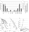
Fig 1
Fig 1. Genetic interactions define a mutation pathway that is dependent upon dinBpolB and nucleotide excision repair genes.
1A: RifR mutation frequencies were determined in various strains in response to UV irradiation. All strains in fig 1A are constructed in the MG1655 background. To account for the intrinsic differences in UV sensitivity among strains, we compared UV doses leading to similar levels of survival: grey bars correspond to UV doses leading to survival levels ranging between 5–15%, for black bars survival levels range between 1–5% survival. It should be stressed that at these UV doses, the SOS response is fully induced in all strains. White bars represent the level of spontaneous mutation frequency, i.e. no UV irradiation. Average values and standard deviations are plotted for three or more independent experiments per strain. 1B: The polA1 allele data are presented in a separate panel as the background in which this allele resides is w3110. Background w3110 exhibits a ≈2-fold higher UV-induced mutagenic response compared to the MG1655 background at UV irradiation levels leading to similar survival. Grey bars correspond to UV doses leading to survival levels ranging between 5–15%; white bars represent the level of spontaneous mutation frequency, i.e. no UV irradiation. 1C: Survival curves of DNA polymerase mutant strains. Wild-type: ■squares; polB: (horizontal segment); umuDC: Δ triangles; dinB:◆ losanges; dinBpolB: X crosses; dinBpolBumuDC: ★ stars. 1D: Survival curves of DNA polymerase mutant strains in an NER-defective background: uvrA •(dots), uvrAdinBpolB ✚ (crosses), uvrAumuDC: (horizontal segment). 1E: UV-induced mutation frequencies plotted as a function of survival in various strains. Wild-type strain ■(squares), uvrA •(dots), dinBpolB X (crosses), uvrAdinBpolB ✚(crosses), umuDC Δ (triangles).
Fig 2
Fig 2. Differential kinetics of mutations induced during NER versus replication.
2A: Kinetics of rifR mutation fixation in various strains following a single UV dose. The UV dose was chosen so as to affect survival of the various strains to a similar extent i.e. ≈10%: wt ■(squares): 100 J/m2, dinBpolB X (crosses): 35 J/m2, uvrA •(dots): 3 J/m2. Following irradiation at the indicated UV dose, cells were platted on rif plates at various time points. RifR mutations accumulate rapidly in the wild-type strain (t1/2 ≈ 30 min after irradiation); in contrast the kinetics of appearance of mutants in the uvrA and in the dinBpolB strains are severely delayed (t1/2 ≈ 80–100 min after irradiation). The plateau reached in the uvrA and dinBpolB strains correspond to ≈ 25–30% and 15–20% of the level reached in the wild-type strain, respectively (see also Fig 1E). 2B: Kinetics of induction of rifR mutation in a dnaBts thermo-sensitive replication mutant irradiated at 90 J/m2 [45,46]. At this dose, survival of dnaBts strain is 11.5%. The dnaBts strain quickly stops replication when shifted at 42°C, the non-permissive temperature. RifR mutants are induced at similar kinetics at permissive (35°C: Δtriangles) and non-permissive (42°C: ■squares) temperature, implying that most mutations are fixed in a non-replicative manner.
Fig 3
Fig 3. Mutations induced during NER and replication: Two hits versus single hit dose response curves (or quadratic versus linear dose response curves).
The induction of rifR mutation frequencies as a function of UV dose in wild-type ■ (squares), dinBpolB X (crosses) and uvrA •(dots) strains (average values of 5 independent experiments per strain). A: In the wild-type strain, the mutation frequency expressed as a function of UV dose, is clearly non-linear. B. The wild-type data fit a straight line when mutation frequencies are plotted as a function of square dose (regression coefficient = 0.96), strongly suggesting that most mutations arise via a two-hit mechanism such as the processing of closely spaced lesions. C and D: In strains dinBpolB (regression coefficient = 0.91) and uvrA (regression coefficient = 0.94) the mutation frequency response is linear with UV dose as expected when mutations occur at the fork during replication.
Fig 4
Fig 4. Opposing lesion structure model for NER-induced mutagenesis.
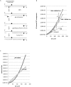
A: Nucleotide Excision Repair initiates repair at a given lesion via its normal dual incision step creating a 12–13 nt single-stranded gap. At rare occasions a second lesion will be located in the opposite strand within the initial incision gap or in close proximity. In the latter case the second lesion may becomes exposed within the gap following a gap enlargement step possibly triggered by the distortion induced by the second lesion at the double-stranded / single-stranded junction via exonuclease (or helicase) processing. The resulting structure will be referred to as “opposing lesion structure”. Alternatively, gap enlargement may result from the necessity to allow a RecA filament to assemble downstream from the lesion in order to activate Pol V [47]. The gap-filling process requires the action of Pol IV/Pol II. The mutation “m” opposite the lesion site is fixed by Pol V during the lesion bypass step. B: Mathematical modeling of UV-induced mutagenesis in a wild-type and dinBpolB strains as a function of UV dose (J/m2). The RiM line fits properly the dinBpolB data points •(dots) (regression coefficient R = 0.91). The wild-type data points ■ (squares) fit appropriately the theoretical curve when the region that defines the opposing lesion zone is set to 18 +/-3 nt (i.e. data points within the 15 and 21 nt range). C: Number of lesions “at risk” (LAR) as a function of UV dose (J/m2). The straight line and the quadratic curve represent the number of lesions, within the rpoB target, at risk for inducing mutations during replication (LAR-RiM) or during NER (LAR-NERiM: opposing lesion zone set to 18nt), respectively (see text). At ≈100J/m2, when the two curves cross, an equal number of mutations will result from replication and NER pathways.
Fig 5
Fig 5. Plasmid-based assay to show NERiM for oxidative lesions.
How general is NERiM? The involvement of NER in induced mutagenesis was investigated by implementation of a plasmid-borne mutation assay using a different mutagenic treatment, namely oxidative damage induced by treating plasmid DNA with Methylene Blue and visible light (MB+ light). A: Experimental outline: the assay involves a pBR322-derived plasmid that contains a +2 frameshift mutation within its tetracycline resistance gene [58]. The assay was shown to monitor true -2 frameshift reversions that restore tetracycline resistacrossnce [59,60]. Plasmid DNA is randomly damaged in vitro with MB+ light treatment and introduced into bacteria by transformation. Cells are plated on tetracycline and ampicillin plates to determine the TetS-> TetR reversion frequency. B: cells are treated by UV irradiation prior to plasmid transformation in order to induce their SOS response. The UV dose is chosen so as to yield a survival of 10–20% (wild-type: 95 J/m2; dinBpolB: 45 J/m2; uvrA: 5 J/m2 and uvrAdinBpolB: 4 J/m2). The average and standard deviation of four to six independent determinations are plotted for each strain. Introduction, into wild-type cells, of plasmid DNA treated with MB+light robustly increases the -2 frameshift mutation frequency by two to three orders of magnitude above untreated control plasmid. The mutagenic response is reduced ≈5-fold when the (MB+light) treated plasmid is introduced into either uvrA, dinBpolB or uvrAdinBpolB strains.
Fig 6
Fig 6. Occurrence of NERiM in stationary phase E. coli cells.
When irradiated at 200 J/m2 in stationary phase the survival of the wt and uvrA strains were equal to 75% and 63%, respectively. A: Induction of rifR mutations by UV-light at a dose of 200 J/m2 (black bar) or in the absence of irradiation (white bar) in stationary phase E. coli cells. B: suppression of UV-induced mutagenesis in stationary E. coli cells that are defective in SOS induction: lexA(Ind) strain.

Similar articles

Cited by

References

    1. Reardon JT, Sancar A. Nucleotide Excision Repair Progress in nucleic acid research and molecular …. Elsevier; 2005. pp. 183–235. doi: 10.1016/S0079-6603(04)79004-2 - DOI - PubMed
    1. Li C-L, Golebiowski FM, Onishi Y, Samara NL, Sugasawa K, Yang W. Tripartite DNA Lesion Recognition and Verification by XPC, TFIIH, and XPA in Nucleotide Excision Repair. Mol Cell. 2015;59: 1025–1034. doi: 10.1016/j.molcel.2015.08.012 - DOI - PMC - PubMed
    1. Staresincic L, Fagbemi AF, Enzlin JH, Gourdin AM, Wijgers N, Dunand-Sauthier I, et al. Coordination of dual incision and repair synthesis in human nucleotide excision repair. EMBO J. 2009;28: 1111–1120. doi: 10.1038/emboj.2009.49 - DOI - PMC - PubMed
    1. Novarina D, Amara F, Lazzaro F, Plevani P, Muzi-Falconi M. Mind the gap: keeping UV lesions in check. DNA Repair (Amst). 2011;10: 751–759. doi: 10.1016/j.dnarep.2011.04.030 - DOI - PMC - PubMed
    1. Howard-Flanders P, Theriot L. Mutants of Escherichia coli K-12 defective in DNA repair and in genetic recombination. Genetics. 1966;53: 1137–1150. - PMC - PubMed

MeSH terms

Substances

LinkOut - more resources