. 2017 Jul 7;12(7):e0180615.
doi: 10.1371/journal.pone.0180615. eCollection 2017.

Therapeutic impact of dietary vitamin D supplementation for preventing right ventricular remodeling and improving survival in pulmonary hypertension

Affiliations

Therapeutic impact of dietary vitamin D supplementation for preventing right ventricular remodeling and improving survival in pulmonary hypertension

Hiroaki Tanaka et al. PLoS One. .

Abstract

Background: Pulmonary hypertension (PH), caused by elevated pulmonary vascular resistance, leads to right heart failure and ultimately death. Vitamin D deficiency can predispose individuals to hypertension and left ventricular dysfunction; however, it remains unknown how serum vitamin D level is related to PH and right ventricular (RV) dysfunction.

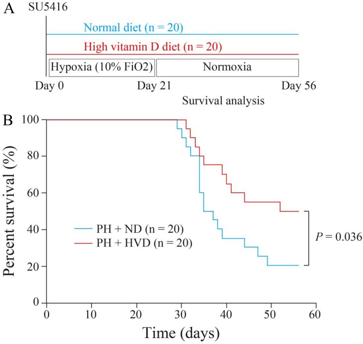
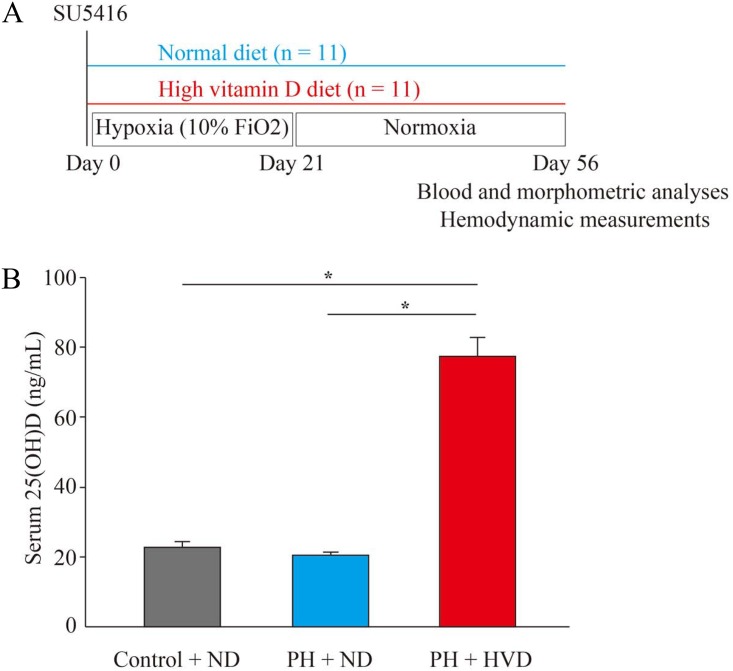
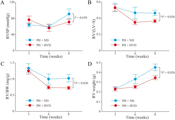
Methods: Serum 25-hydroxyvitamin D [25(OH)D] levels were assessed in PH patients for an association with disease severity. To examine whether vitamin D supplementation could prevent the development of pulmonary vascular remodeling and RV dysfunction in PH, a rat model of PH was fed either normal chow or a high vitamin D diet.

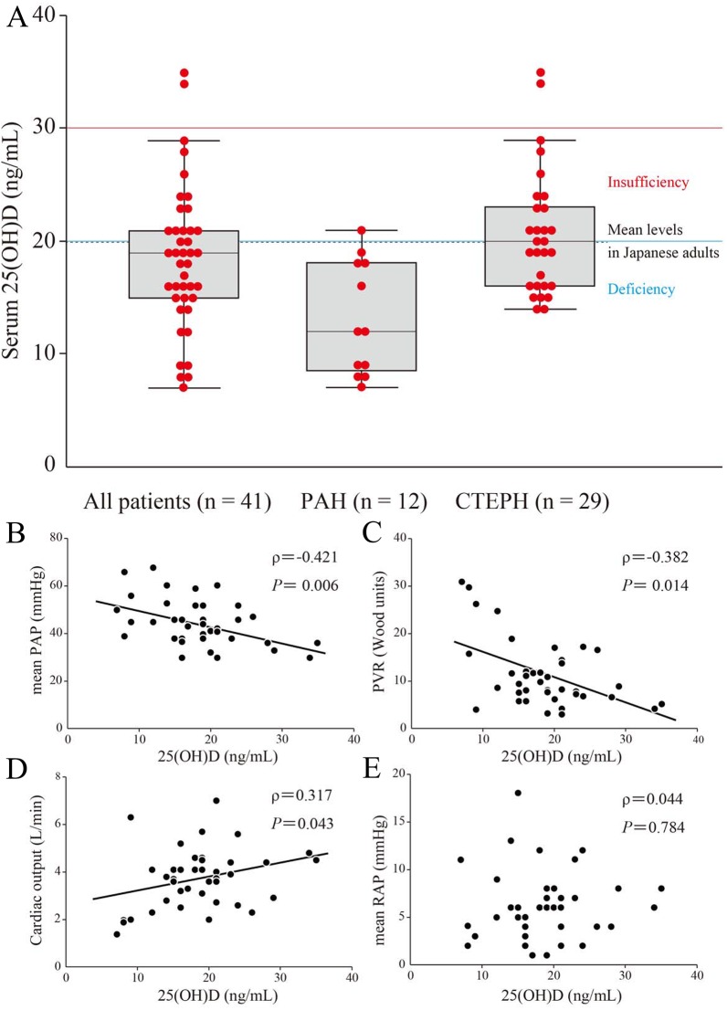
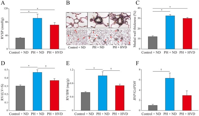
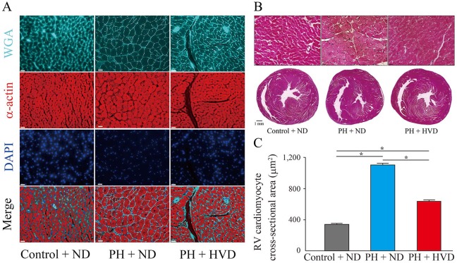
Results: The majority (95.1%) of PH patients had 25(OH)D levels in the insufficiency range, which is associated with increased mean pulmonary artery pressure, increased pulmonary vascular resistance, and decreased cardiac output in PH patients. Vitamin D supplementation significantly increased serum 25(OH)D levels and improved survival in PH rats. Interestingly, while the supplemented rats retained the typical increases in medial thickness of the muscular pulmonary arteries and RV systolic pressure, RV cardiomyocyte hypertrophy and B-type natriuretic peptide expression was significantly attenuated.

Conclusions: Vitamin D deficiency is frequently seen in patients diagnosed with PH and low serum levels of 25(OH)D are associated with severity of PH and RV dysfunction. Vitamin D supplementation in PH rats improved survival via ameliorating pathological RV hypertrophy. These findings suggest an insufficient intake of vitamin D might potentially accelerate RV dysfunction, leading to a crucial clinical impact of vitamin D supplementation in PH.


Conflict of interest statement

Competing Interests: Actelion Pharmaceuticals Japan Ltd. provided support in the form of salaries for an author [HT]. This does not alter our adherence to PLOS ONE policies on sharing data and materials.

Figures

Fig 1
Fig 1. Relationship between serum 25-hydroxyvitamin D (25(OH)D) levels and hemodynamic parameters in patients with pulmonary hypertension (PH).
Serum 25(OH)D levels and hemodynamic parameters were measured in 41 patients with PH, consisting of 12 patients with idiopathic or heritable pulmonary arterial hypertension (PAH) and 29 patients with chronic thromboembolic pulmonary hypertension (CTEPH). (A) Serum 25(OH)D levels in each PH classification. Serum 25(OH)D levels are shown as box plots, with bars denoting median values, boxes denoting interquartile ranges, and whiskers denoting ranges, excluding statistical outliers (●; >1.5 box lengths from either the 25th or 75th percentiles). Serum 25(OH)D levels of insufficiency (red line), deficiency (blue line), and the mean levels in Japanese adults (black broken line) are shown. (B)-(E) Correlations in 41 patients with PH were analyzed between 25(OH)D levels and mean pulmonary arterial pressure (PAP) (B), pulmonary vascular resistance (PVR) (C), cardiac output (D), and mean right atrial pressure (RAP) (E), respectively.
Fig 2
Fig 2. Survival benefit of dietary vitamin D supplementation in PH rats.
(A) Experimental protocol for investigating the effects of dietary vitamin D supplementation on survival in Fisher 344 rats with PH. (B) Kaplan-Meier survival curves of PH rats receiving the normal diet (PH + ND group) and PH rats receiving the high vitamin D diet (PH + HVD group) (n = 20 rats per group).
Fig 3
Fig 3. Experimental protocol and serum 25(OH)D levels in PH rats.
(A) Experimental protocol for investigating the effects of dietary vitamin D supplementation in Sprague-Dawley rats with PH. (B) Serum 25(OH)D levels in control rats receiving the normal diet (control + ND group), PH rats receiving the normal diet (PH + ND group), or PH rats receiving the high vitamin D diet (PH + HVD group) (n = 8 rats per group). Data are mean ± SEM. *P < 0.01.
Fig 4
Fig 4. Effects of dietary vitamin D supplementation in PH rats.
Assessments in Control + ND group, PH + ND group, and PH + HVD group (n = 10–11 rats per group). (A) Right ventricular systolic pressure (RVSP). (B) Representative Elastica-van Gieson (EVG) staining of lungs from rats in each experimental group. Red arrows indicate pulmonary arteries. Scale bars, 20 μm (above panels) and 100 μm (below panels). (C) Percent medial wall thickness in the small pulmonary arteries of 20–100 μm diameter (n = 4–10 rats per group). (D) The ratio of right ventricular weight to left ventricle plus septum weight [RV/(LV+S)]. (E) The ratio of right ventricular weight to body weight (RV/BW). (F) Relative expression of B-type natriuretic peptide (BNP) mRNA levels normalized to levels of glyceraldehyde 3-phosphate dehydrogenase (GAPDH) in the right ventricles of experimental rats. Expression was quantified by qPCR (n = 3–4 rats per group). Data are mean ± SEM. *P < 0.01, †P < 0.05.
Fig 5
Fig 5. Inhibition of hypertrophy in right ventricular (RV) cardiomyocytes by dietary vitamin D supplementation in PH rats.
(A) and (B), Representative images of hearts from rats in each experimental group. (A), Wheat germ agglutinin (WGA), α-actin, and 4’,6-diamidino-2-phenylindole (DAPI) staining of RV cross sections. Scale bars, 20 μm. (B) Hematoxylin and eosin (H&E) staining. Above panels show magnified images of cardiomyocytes in RV wall. Scale bars, 20 μm (above panels) and 1 mm (below panels). (C) Quantification of RV cardiomyocyte with cross-sectional area stained (n = 5 rats per group). Data are mean ± SEM. *P < 0.01.
Fig 6
Fig 6. Time-course changes of PH and RV remodeling in PH rats.
Time-course data were compared between PH + ND group and PH + HVD group (n = 5–11 rats per group). (A) RV systolic pressure (RVSP). (B) The ratio of right ventricular weight to left ventricle plus septum weight [RV/(LV+S)]. (C) The ratio of right ventricular weight to body weight (RV/BW). (D) RV weight. Data are mean ± SEM. *P < 0.05.

References

    1. Pulido T, Adzerikho I, Channick RN, Delcroix M, Galiè N, Ghofrani HA, et al. Macitentan and morbidity and mortality in pulmonary arterial hypertension. N Engl J Med. 2013;369:809–18. doi: 10.1056/NEJMoa1213917 - DOI - PubMed
    1. Sitbon O, Channick R, Chin KM, Frey A, Gaine S, Galiè N, et al. Selexipag for the treatment of pulmonary arterial hypertension. N Engl J Med. 2015;373:2522–33. doi: 10.1056/NEJMoa1503184 - DOI - PubMed
    1. Farber HW, Miller DP, Poms AD, Badesch DB, Frost AE, Muros-Le Rouzic E, et al. Chest. 2015;148:1043–54. doi: 10.1378/chest.15-0300 - DOI - PubMed
    1. Delcroix M, Lang I, Pepke-Zaba J, Jansa P, D'Armini AM, Snijder R, et al. Long-term outcome of patients with chronic thromboembolic pulmonary hypertension: results from an international prospective registry. Circulation. 2016;133:859–71. doi: 10.1161/CIRCULATIONAHA.115.016522 - DOI - PubMed
    1. Holick MF. Vitamin D deficiency. N Engl J Med. 2007;357:266–81. doi: 10.1056/NEJMra070553 - DOI - PubMed

MeSH terms