. 2017 Jul 7;12(7):e0180981.
doi: 10.1371/journal.pone.0180981. eCollection 2017.

Plasminogen Tochigi mice exhibit phenotypes similar to wild-type mice under experimental thrombotic conditions

Affiliations

Plasminogen Tochigi mice exhibit phenotypes similar to wild-type mice under experimental thrombotic conditions

Yuko Tashima et al. PLoS One. .

Abstract

Plasminogen (Plg) is a precursor of plasmin that degrades fibrin. A race-specific A620T mutation in Plg, also known as Plg-Tochigi, originally identified in a patient with recurrent venous thromboembolism, causes dysplasminogenemia with reduced plasmin activity. The Plg-A620T mutation is present in 3-4% of individuals in East Asian populations, and as many as 50,000 Japanese are estimated to be homozygous for the mutant 620T allele. In the present study, to understand the changes of thrombotic phenotypes in individuals with the mutant 620T allele, we generated knock-in mice carrying the homozygous Plg-A622T mutation (PlgT/T), an equivalent to the A620T mutation in human Plg. PlgT/T mice grew normally but showed severely reduced plasmin activity activated by urokinase, equivalent to ~8% of that in wild-type mice. In vitro fibrin clot lysis in plasma was significantly slower in PlgT/T mice than in wild-type mice. However, all experimental models of electrolytic deep vein thrombosis, tissue factor-induced pulmonary embolism, transient focal brain ischaemic stroke, or skin-wound healing showed largely similar phenotypes between PlgT/T mice and wild-type mice. Protein S-K196E mutation (Pros1E/E) is a race-specific genetic risk factor for venous thromboembolism. Coexistence in mice of PlgT/T and Pros1E/E did not affect pulmonary embolism symptoms, compared with those in Pros1E/E mice. Hence, the present study showed that the Plg-A622T mutation, which confers ~8% plasmin activity, does not increase the risk of thrombotic diseases in mice under experimental thrombotic conditions and does not modify the thrombotic phenotype observed in Pros1E/E mice. PlgT/T mice can be used to investigate the potential pathophysiological impact of the Plg-A620T mutation.


Conflict of interest statement

Competing Interests: The authors have declared that no competing interests exist.

Figures

Fig 1
Fig 1. Generation of Plg-A622T mice.
(A) Structure of the targeted locus in the mouse Plg gene. Exons are represented by filled boxes. A loxP-flanked (filled triangles) neomycin-resistance cassette (NEO) and a diphtheria toxin A fragment expression cassette (DT-A) are indicated by open boxes with arrows that represent the transcriptional orientation. The A622T mutant allele was produced by homologous recombination and NEO deletion mediated by Cre recombinase. The c.1864G>A (p.A622T) mutation and three translationally silent mutations (c.1857T>G, c.1858C>T, c.1860G>A) creating a new HpaI site (GTTAAC) were introduced into exon 15. Homologous fragments are indicated by dotted lines, while the MfeI-HpaI fragments detected by Southern blot analysis of the wild-type (WT) and Plg-A622T alleles are indicated by double-headed arrows. (B) Southern blot analysis. Genomic DNA from targeted ES cells was digested with MfeI/HpaI and detected with the specific probe (WT allele: 9.5 kb; Plg-A622T allele: 6.6 kb). (C) Quantitative RT-PCR analysis. Total RNA was extracted from mouse liver and subjected to real-time RT-PCR with dual-labeled probes for mouse Plg and Rn18s. Expression levels of Plg mRNA were normalized to Rn18s mRNA. Data are the means ± SDs of WT (n = 8) and PlgT/T (n = 7) mice. The levels measured in WT mice were defined as 100%.
Fig 2
Fig 2. Plasma Plg antigen levels and plasmin activities of wild-type and PlgT/T mice.
(A) Plg antigen levels. Data are the means ± SDs of wild-type (WT, n = 10) and PlgT/T (n = 10) mice. The levels measured in WT mice were defined as 1 U/ml. (B) Plasmin activities. Plasma from the indicated mice was preincubated with human uPA and reacted with a synthetic substrate, S-2403, for plasmin. Data are the means ± SDs of 10 mice for each genotype. The mean activity measured in WT mice was defined as 100%. (C) Western blot analysis of uPA-treated mouse plasma. Two mouse plasma samples of each genotype were incubated with human uPA and separated by SDS-PAGE under reducing conditions. Plg (black triangle) and heavy chains of plasmin (white triangles) were detected with anti-mouse Plg antibodies.
Fig 3
Fig 3. Effect of Plg-A622T mutation on fibrinolysis in plasma.
Pooled plasma from 6 wild-type (A) or 6 PlgT/T (B) mice was preincubated in the absence (○) or presence (◆) of human tPA, and clotting was induced with thrombin and CaCl2. Human α2-plasmin inhibitor (α2-PI) was added before the induction of clotting (▽). The turbidity monitored by the absorbance at 405 nm was measured every 5 min as an index of fibrin formation and lysis. The time course of the turbidity was plotted to the maximum absorbance of each sample, which was taken as 100%.
Fig 4
Fig 4. Thrombus weights in electrolytic IVC injury-induced DVT in wild-type and PlgT/T mice.
On day 2 or day 7 post-injury, thrombi formed in the IVC were weighed. No significant differences (p > 0.05) were observed in the thrombus weight between wild-type (WT) and PlgT/T mice (n = 9 and n = 9 on day 2, and n = 5 and n = 5 on day 7, respectively). Circles and squares represent individual mouse data. Bars represent the mean values of groups.
Fig 5
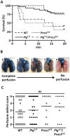
Fig 5. Survival time and lung perfusion defect after tissue factor-induced PE.
(A) After the tissue factor infusion via IVC, the survival time was recorded until 20 min. No significant difference was observed in the survival between wild-type (WT) (n = 28) and PlgT/T (n = 29) mice (hazard ratio, 1.18; 95% confidence intervals, 0.40–3.50), or between Pros1E/E (n = 10) and PlgT/T/Pros1E/E (n = 10) mice (hazard ratio, 0.81; 95% confidence intervals, 0.27–2.41). The survival rates of both WT and PlgT/T mice were significantly longer than those of Pros1E/E and PlgT/T/Pros1E/E mice (WT and Pros1E/E mice: hazard ratio, 5.43; 95% confidence intervals, 1.81–16.28; WT and PlgT/T/Pros1E/E mice: hazard ratio, 4.38; 95% confidence intervals, 1.41–13.60; PlgT/T and Pros1E/E mice: hazard ratio, 4.62; 95% confidence intervals, 1.61–13.28; PlgT/T and PlgT/T/Pros1E/E mice: hazard ratio, 3.73; 95% confidence intervals, 1.25–11.11). *Significant difference in comparison to WT mice and PlgT/T mice. (B) The scale used to measure lung perfusion defect scores. A score of 0 indicates complete perfusion of Evans blue with no occlusion and a score of 4 indicates no Evans blue perfusion with complete occlusion. (C) Perfusion defect scores. No significant (ns) differences (p > 0.05) were observed among WT (n = 28), PlgT/T (n = 29), Pros1E/E (n = 10) and PlgT/T/Pros1E/E mice (n = 10). Perfusion defect scores were assessed by the Mann-Whitney test. Circles and diamonds represent individual mouse data. Bars represent the mean values of groups.
Fig 6
Fig 6. No exacerbation of PlgT/T mutation in the transient focal brain ischaemia model.
(A) Representative images of coronal sections of wild-type (WT) and PlgT/T mouse brains. Permanent occlusion of the distal M1 portion of the left middle cerebral artery and 15-min transient occlusion of the bilateral common carotid arteries were applied. After 24 hours, the brains were excised and stained with 2, 3, 5-triphenyl tetrazolium chloride. White areas represent brain infarction. (B) Infarct volumes. The infarct volume was adjusted for edema by dividing the volume by the edema index (left hemisphere volume / right hemisphere volume). No significant differences (p > 0.05) were observed between groups. Circles represent individual mouse data. Bars represent the mean values of groups.
Fig 7
Fig 7. No effects of PlgT/T mutation in the skin-wound healing model.
The time course of the wound area was measured every other day for two weeks. Data are the means ± SDs of wild-type (WT, n = 10) and PlgT/T (n = 9) mice. No significant differences (p > 0.05) were observed between groups.

Similar articles

Cited by

References

    1. Castellino FJ, Ploplis VA. Structure and function of the plasminogen/plasmin system. Thromb Haemost. 2005;93(4):647–54. doi: 10.1160/TH04-12-0842 . - DOI - PubMed
    1. Schuster V, Hugle B, Tefs K. Plasminogen deficiency. J Thromb Haemost. 2007;5(12):2315–22. doi: 10.1111/j.1538-7836.2007.02776.x . - DOI - PubMed
    1. Rijken DC, Lijnen HR. New insights into the molecular mechanisms of the fibrinolytic system. J Thromb Haemost. 2009;7(1):4–13. doi: 10.1111/j.1538-7836.2008.03220.x . - DOI - PubMed
    1. Schuster V, Mingers AM, Seidenspinner S, Nussgens Z, Pukrop T, Kreth HW. Homozygous mutations in the plasminogen gene of two unrelated girls with ligneous conjunctivitis. Blood. 1997;90(3):958–66. . - PubMed
    1. Tefs K, Gueorguieva M, Klammt J, Allen CM, Aktas D, Anlar FY, et al. Molecular and clinical spectrum of type I plasminogen deficiency: A series of 50 patients. Blood. 2006;108(9):3021–6. doi: 10.1182/blood-2006-04-017350 . - DOI - PubMed

MeSH terms

Supplementary concepts