. 2017 Jul 6;101(1):87-103.
doi: 10.1016/j.ajhg.2017.06.007.

A Pentanucleotide ATTTC Repeat Insertion in the Non-coding Region of DAB1, Mapping to SCA37, Causes Spinocerebellar Ataxia

Affiliations

A Pentanucleotide ATTTC Repeat Insertion in the Non-coding Region of DAB1, Mapping to SCA37, Causes Spinocerebellar Ataxia

Ana I Seixas et al. Am J Hum Genet. .

Abstract

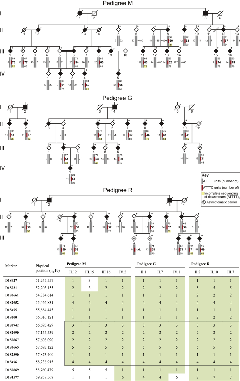
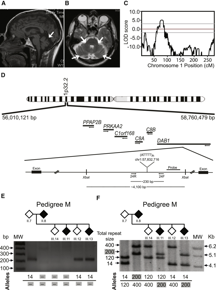
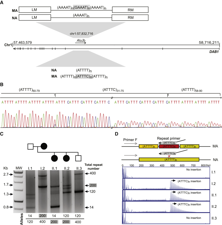
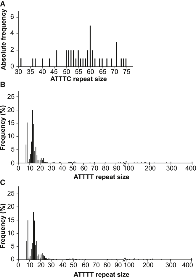
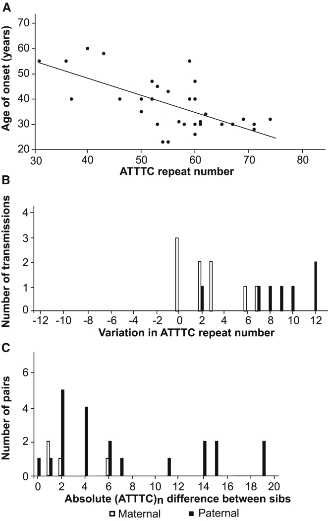
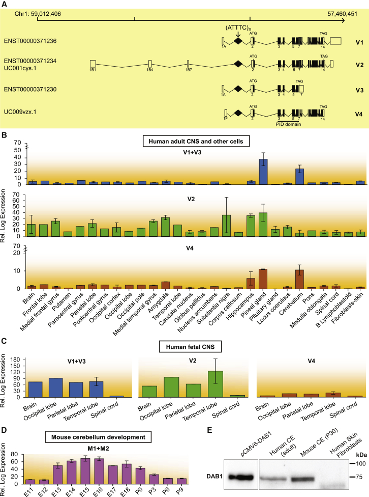
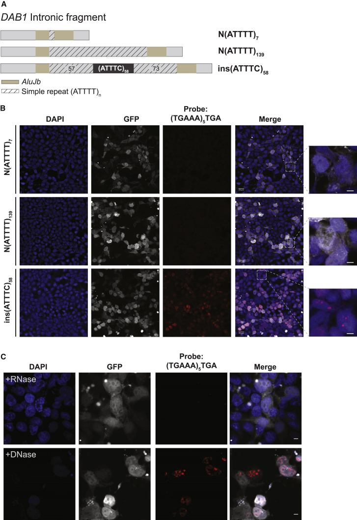
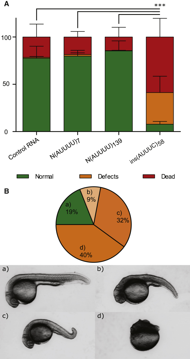
Advances in human genetics in recent years have largely been driven by next-generation sequencing (NGS); however, the discovery of disease-related gene mutations has been biased toward the exome because the large and very repetitive regions that characterize the non-coding genome remain difficult to reach by that technology. For autosomal-dominant spinocerebellar ataxias (SCAs), 28 genes have been identified, but only five SCAs originate from non-coding mutations. Over half of SCA-affected families, however, remain without a genetic diagnosis. We used genome-wide linkage analysis, NGS, and repeat analysis to identify an (ATTTC)n insertion in a polymorphic ATTTT repeat in DAB1 in chromosomal region 1p32.2 as the cause of autosomal-dominant SCA; this region has been previously linked to SCA37. The non-pathogenic and pathogenic alleles have the configurations [(ATTTT)7-400] and [(ATTTT)60-79(ATTTC)31-75(ATTTT)58-90], respectively. (ATTTC)n insertions are present on a distinct haplotype and show an inverse correlation between size and age of onset. In the DAB1-oriented strand, (ATTTC)n is located in 5' UTR introns of cerebellar-specific transcripts arising mostly during human fetal brain development from the usage of alternative promoters, but it is maintained in the adult cerebellum. Overexpression of the transfected (ATTTC)58 insertion, but not (ATTTT)n, leads to abnormal nuclear RNA accumulation. Zebrafish embryos injected with RNA of the (AUUUC)58 insertion, but not (AUUUU)n, showed lethal developmental malformations. Together, these results establish an unstable repeat insertion in DAB1 as a cause of cerebellar degeneration; on the basis of the genetic and phenotypic evidence, we propose this mutation as the molecular basis for SCA37.

Keywords: DAB1 reelin adaptor protein; RNA-mediated toxicity; SCA37; large Alu pentanucleotide repeat; neurodegeneration; neurodegenerative disease; neurodevelopmental gene; repeat expansion; repeat instability; unstable repeat insertion.


Figures

Figure 1
Figure 1
Pedigrees of SCA-Affected Families and STR Haplotypes Pedigree structures of the three families used for genome-wide linkage analysis and the corresponding individual genotypes and pentanucleotide repeat configuration. Below are the disease haplotypes from selected affected individuals, identified by pedigree number, for 14 markers from locus D1S427 (51,245,557 bp) to D1S1577 (59,958,568). The disease haplotypes found in the families established an initial candidate region spanning 2.8 Mb between markers D1S200 and D1S2869 in chromosomal region 1p32.2 (hg19) (shown in a box). Symbols in pedigrees were modified for privacy protection. Abbreviations are as follows: n.d., not determined; and +, individual for whom DNA was available.
Figure 2
Figure 2
Chromosome Mapping and Mutation Screening (A and B) Brain MRI of affected individual M8 (Table 1) shows atrophy of (A) the cerebellar cortex on sagittal T1 and (B) the middle cerebellar peduncles on an axial T2-weighted image. (C) Linkage analysis using genotypes from 18 individuals from family M shows a maximum multipoint LOD score of 5.1 obtained in chromosomal region 1p32. (D) Schematic physical map of the SCA candidate region; also depicted are the intronic repeat region with the XbaI restriction sites, the location of the Southern blot probe, and primers used for standard PCR. The (ATTTT)n position according to hg19 is shown. (E) Standard PCR analysis of the pentanucleotide repeat ATTTT/AAAAT in the intronic 5′ UTR of DAB1 shows a lack of PCR amplification of large repeat tracts that were not amplified by standard PCR. (F) Using a probe hybridizing near the pentanucleotide repeat ATTTT/AAAAT in DAB1, corresponding Southern blot analysis for the same family branch in (E) shows that an ∼5.1-kb fragment corresponding to ∼200 pentanucleotide repeats was transmitted from the affected mother to affected offspring. Large non-pathogenic (ATTTT)n alleles are unstable, and the estimated allele size of 120 on Southern blot was shown by sequencing analysis to vary from 127 to 139 units. Individual ID numbers are the same as those in each pedigree in Figure 1.
Figure 3
Figure 3
Identification of an Unstable ATTTC Repeat Insertion in DAB1 (A) Schematic representation of the ATTTT/AAAAT simple repeat flanked by left (LM) and right (RM) monomers in the polymorphic middle A-rich region of an AluJb sequence in an intron of the DAB1 5′ UTR; also depicted is the structure of normal alleles (NAs) with pure ATTTT/AAAAT repeats and mutant alleles (MAs) with the (ATTTC)n insertion. (B) Sequencing analysis of the DAB1 repeat shows the (ATTTC)n insertion in the middle of the ATTTT simple repeat in affected individuals. (C) Long-range PCR across the Alu sequence was used for assaying alleles up to 2.7 kb. (D) In this family branch shown for long-range PCR, the ATTTC repeat insertion was readily detected by RP-PCR analysis in affected individuals (mother I.2 and offspring II.1 and II.2) but not in the unaffected individuals I.1 and II.3.
Figure 4
Figure 4
Distribution of Pathological and Normal Alleles (A) The absolute frequency of pathological (ATTTC)n allele sizes in SCA subjects (n = 41). (B and C) Distribution of (ATTTT)n allele sizes in chromosomes from (B) the normal population (n = 520) and (C) affected individuals with other neurodegenerative diseases (n = 904).
Figure 5
Figure 5
Inverse Correlation between (ATTTC)n Size and Age of Onset in SCA Subjects and Instability (A) A scatterplot showing an inverse correlation between the length of the (ATTTC)n insertion and age of onset. Affected individuals with larger insertion sizes present with earlier onset (r = −0.68, p < 0.001, n = 33). (B) Parent-offspring transmissions with varying ATTTC repeat numbers with paternal or maternal origin. (C) Difference in ATTTC insertion size in pairs of siblings upon paternal and maternal transmissions.
Figure 6
Figure 6
Expression of DAB1 Transcripts Spanning the Region Containing the Repeat Insertion and DAB1 (A) Schematics of DAB1 genomic position on chromosome 1 show transcripts identified by CAGE (FAMTOM5) to result from usage of alternative promoters. These transcripts are also annotated in Ensembl or the UCSC Genome Browser (hg19), and all have the repeat insertion region in the 5′ UTR. Coding and non-coding exons in transcripts are represented by closed and open boxes, respectively. The location of the (ATTTC)n insertion region in transcript variants is represented by a diamond and indicated with an arrow, and the locations of the ATG start codon and TAG stop codon are shown. The following abbreviation is used: PID, phosphotyrosine interaction domain. (B–D) Mean expression levels of DAB1 transcripts in different CNS regions, skin fibroblasts, and B lymphoblastoid cells from human adults, as analyzed from CAGE data (B); mean expression levels of DAB1 transcript variants in CNS regions of 20- to 29-week-old human fetuses (C); and mean developmental expression levels of Dab1 transcripts in mouse cerebellum from embryonic day 11 (E11) to postnatal day 9 (P9), as analyzed from CAGE data (D). Samples available for expression analysis of each CNS region or cell type ranged from one to three in human adults and from one to two in human fetuses, whereas in mouse cerebellum, three samples were available for all stages. Data represent the mean ± SD. (E) Western blot analysis of DAB1 localization in adult human and mouse cerebellum and in primary skin fibroblasts. DAB1 from HEK293T cells overexpressing human DAB1 cDNA plasmid (pCMV6-hDAB1, OriGene Technologies) was used as a control. Boxes indicate cropped areas from digital images of the same membrane obtained at different exposure times (30 s for the first lane and 5 min for the other three lanes).
Figure 7
Figure 7
Formation of (AUUUC)n RNA Aggregates in a Human Cell Line (A) Two normal Alu fragments with a simple ATTTT repeat of different size and the pathogenic ATTTC repeat insert (with the configuration (ATTTT)57(ATTTC)58(ATTTT)73) were directly amplified from genomic DNA and cloned into the pCDH-CMV-EF1-GFP-Puro vector. (B) Transient overexpression of the pathogenic ATTTC repeat insertion, but not the normal ATTTT repeat of 7 or 139 units, in HEK293T cells leads to widespread formation of nuclear RNA aggregates visible after FISH staining with a probe, (TGAAA)5TGA, predicted to hybridize to (AUUUC)n. GFP expression was used as a marker for transfection. Represented are single-plane confocal images. Scale bars, 5 μm. (C) Aggregates, variable in size and number in individual cells, were sensitive to enzymatic treatment with RNase but not DNase. Representative images are projections of z stacked images collected at 0.21 μm intervals with a confocal microscope. Scale bars, 5 μm.
Figure 8
Figure 8
In Vivo Deleterious Effects of the (ATTTC)n Insertion (A) Percentage of embryos that presented with lethality (dead), developmental defects (defects), or a wild-type phenotype (normal) at 24 hpf after RNA injection of control Cas9RNA, N(AUUUU)7, N(AUUUU)139, or ins(AUUUC)58 (average of three replicas with at least 200 embryos per replica; ∗∗∗p ≤ 0.001; χ2 test for the lethality rate). Data represent the mean ± SD. (B) Distribution of phenotypic classes (a–d) observed in ins(AUUUC)58-injected embryos at 24 hpf and representative images of the observed phenotypic classes: (a) wild-type, (b) severe defects in the tail and head, (c) mild defect in the tail, and (d) severe defects in the anterior-posterior axis.

References

    1. Sequeiros J., Martins S., Silveira I. Epidemiology and population genetics of degenerative ataxias. Handb. Clin. Neurol. 2012;103:227–251. - PubMed
    1. Durr A. Autosomal dominant cerebellar ataxias: polyglutamine expansions and beyond. Lancet Neurol. 2010;9:885–894. - PubMed
    1. Coutinho P., Ruano L., Loureiro J.L., Cruz V.T., Barros J., Tuna A., Barbot C., Guimarães J., Alonso I., Silveira I. Hereditary ataxia and spastic paraplegia in Portugal: a population-based prevalence study. JAMA Neurol. 2013;70:746–755. - PubMed
    1. Silveira I., Miranda C., Guimarães L., Moreira M.C., Alonso I., Mendonça P., Ferro A., Pinto-Basto J., Coelho J., Ferreirinha F. Trinucleotide repeats in 202 families with ataxia: a small expanded (CAG)n allele at the SCA17 locus. Arch. Neurol. 2002;59:623–629. - PubMed
    1. Carlson K.M., Andresen J.M., Orr H.T. Emerging pathogenic pathways in the spinocerebellar ataxias. Curr. Opin. Genet. Dev. 2009;19:247–253. - PMC - PubMed

MeSH terms