

Recurrence of visceral and muco-cutaneous leishmaniasis in a patient under immunosuppressive therapy
- PMID: 28687071
- PMCID: PMC5501116
- DOI: 10.1186/s12879-017-2571-x
Recurrence of visceral and muco-cutaneous leishmaniasis in a patient under immunosuppressive therapy
Abstract
Background: Leishmaniasis is a protozoan disease caused by parasites of the genus Leishmania, transmitted to humans by sandflies. The diagnosis of leishmaniasis is often challenging as it mimics many other infectious or malignant diseases. The disease can present in three ways: cutaneous, mucocutaneous, or visceral leishmaniasis, which rarely occur together or consecutively.
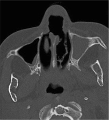
Case presentation: The patient was a 52 years old immunosuppressed Belgian woman with a long history of severe rheumatoid arthritis. She underwent bone marrow biopsy to explore thrombocytopenia. Diagnosis of visceral leishmaniasis was made by identification of Leishman Donovan (LD) bodies in macrophages. Treatment with liposomal amphotericin B was successful. She later developed cutaneous leishmaniasis treated with amphotericin B lipid complex. She next presented with relapsing cutaneous lesions followed by rapidly progressing lymphadenopathies. Biopsy confirmed the diagnosis of leishmaniasis. Treatments by miltefosine, amphotericin B, N-methyl-glucamine antimoniate were subsequently initiated. She later presented a recurrent bone marrow involvement treated with intramuscular paromomycin and miltefosine. She died two years later from leukemia. At the time of death, she presented with a mucosal destruction of the nose. A Leishmania-specific PCR (Polymerase Chain Reaction) identified L. infantum as etiological agent.
Conclusions: Clinicians should be aware of the potential concomitant or sequential involvement of multiple anatomic localizations of Leishmania in immunosuppressed patients.
Keywords: Cutaneous leishmaniasis; Immunosuppression; Microbiology; Mucosal leishmaniasis; Parasitology; Visceral leishmaniasis.
Conflict of interest statement
Ethics approval and consent to participate
Not Applicable.
Consent for publication
Consent to publish was obtained from a next-of-kin after the patient’s death.
Competing interests
The authors declare that they have no competing interests.
Publisher’s Note
Springer Nature remains neutral with regard to jurisdictional claims in published maps and institutional affiliations.
Figures

References
Publication types
MeSH terms
Substances
LinkOut - more resources
Full Text Sources
Other Literature Sources
Research Materials
