

A rare case of malignant solitary fibrous tumor in prostate with review of the literature
- PMID: 28687087
- PMCID: PMC5501453
- DOI: 10.1186/s13000-017-0640-5
A rare case of malignant solitary fibrous tumor in prostate with review of the literature
Abstract
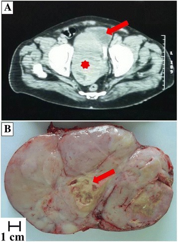
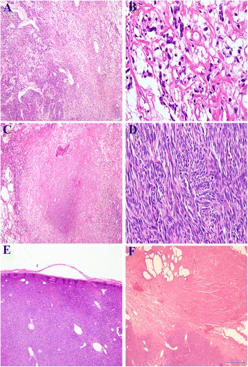
Background: Solitary fibrous tumor is an uncommon soft tissue neoplasm with intermediate biological behavior, which rarely metastasizes. Malignant solitary fibrous tumor, although not clearly defined, is rarely described in the prostate. The present case is characterized by some peculiarities if compared with previously reported cases of prostatic malignant solitary fibrous tumor. Firstly, it does not show a homogeneous morphology: part of the neoplasm (about 50%) showed the features of a conventional solitary fibrous tumor, while the remaining part showed the features of a malignant solitary fibrous tumor. In addition, the case is the first malignant solitary fibrous tumor reaching a huge diameter of 20 cm and replacing all prostatic parenchyma. Interestingly, normal prostatic parenchyma was observed on left-lobe trans-rectal needle-core biopsies, but was totally absent in surgical specimen. Since radical prostatectomy was carried out about 4 months after the biopsies, such discordant data may suggest exceedingly rapid growth of the neoplasm.
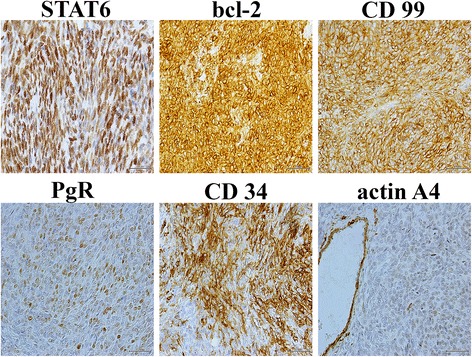
Case presentation: We report a case of a 62-year-old male, presented at medical observation for urinary retention, constipation and an enlarged prostate gland. A trans-rectal prostatic biopsy showed a low-grade spindle cell neoplasm. Histopathological examination of the prostatectomy specimen showed patternless architecture with hypocellular and hypercellular areas and hemangiopericytoma-like vessels. In some fields the neoplasm was characterized by a high mitotic index and evident cellular atypia. Immunohistochemically, neoplastic cells were positive for CD34, bcl2, CD99, STAT6 and partially for PgR. The neoplasm was diagnosed as a malignant solitary fibrous tumor.
Conclusions: The differential diagnosis of spindle cells tumors arising in the prostrate is broad and includes lesions of epithelial and mesenchymal origin, primary prostatic lesions such as stromal tumors of uncertain malignant potential and stromal sarcoma, as well as anatomically ubiquitous soft tissue neoplasms. Solitary fibrous tumors should be considered in cases of prostatic tumors with a spindled morphology, but malignancy in such tumors is extremely rare in the prostate. A review of literature showed only four additional cases. Because of the unpredictable biological behavior and the possibility of recurrence, a long-term clinical and instrumental follow-up is recommended.
Keywords: Haemangiopericytoma; Mesenchymal neoplasm; Prostate neoplasm; Soft tissue tumor; Spindle cell tumor.
Conflict of interest statement
Ethics approval and consent to participate
Not applicable.
Consent for publication
Written informed consent was obtained from the patient for publication of this case report and any accompanying images. A copy of the written consent is available for review by the Editor-in-Chief of this journal.
Competing interests
The authors declare that they have no competing interests.
Publisher’s Note
Springer Nature remains neutral with regard to jurisdictional claims in published maps and institutional affiliations.
Figures
Similar articles
-
Clinicopathological features to distinguish malignant solitary fibrous tumors of the prostate from prostatic stromal tumors.Virchows Arch. 2021 Apr;478(4):619-626. doi: 10.1007/s00428-020-02909-2. Epub 2020 Aug 20. Virchows Arch. 2021. PMID: 32820389
-
Orbital solitary fibrous tumor: encompassing terminology for hemangiopericytoma, giant cell angiofibroma, and fibrous histiocytoma of the orbit: reappraisal of 41 cases.Hum Pathol. 2011 Jan;42(1):120-8. doi: 10.1016/j.humpath.2010.05.021. Epub 2010 Nov 5. Hum Pathol. 2011. PMID: 21056898
-
Primary solitary fibrous tumour of the prostate: A case report and literature review.Malays J Pathol. 2020 Dec;42(3):449-453. Malays J Pathol. 2020. PMID: 33361728 Review.
-
Solitary fibrous tumor on needle biopsy and transurethral resection of the prostate: a clinicopathologic study of 13 cases.Am J Surg Pathol. 2007 Jun;31(6):870-6. doi: 10.1097/01.pas.0000213416.23256.71. Am J Surg Pathol. 2007. PMID: 17527073
-
Massive malignant solitary fibrous tumor arising from the bladder serosa: a case report.J Med Case Rep. 2015 Mar 1;9:46. doi: 10.1186/s13256-014-0505-4. J Med Case Rep. 2015. PMID: 25884588 Free PMC article. Review.
Cited by
-
Primary Prostatic Stromal Sarcoma: A Case Report and Review of the Literature.Medicina (Kaunas). 2024 Nov 22;60(12):1918. doi: 10.3390/medicina60121918. Medicina (Kaunas). 2024. PMID: 39768800 Free PMC article. Review.
-
Case report: 125I seed implantation for rare malignant solitary fibrous tumor in the pelvic cavity: a case report.Front Oncol. 2022 Aug 1;12:884491. doi: 10.3389/fonc.2022.884491. eCollection 2022. Front Oncol. 2022. PMID: 35978802 Free PMC article.
-
A Novel Cause of Bowel Obstruction in a Patient with Long-Standing Crohn's Disease.Case Rep Pathol. 2021 Oct 18;2021:3278392. doi: 10.1155/2021/3278392. eCollection 2021. Case Rep Pathol. 2021. PMID: 34707913 Free PMC article.
-
Intracranial malignant solitary fibrous tumor metastasized to the chest wall: A case report and review of literature.World J Clin Cases. 2020 Oct 26;8(20):4844-4852. doi: 10.12998/wjcc.v8.i20.4844. World J Clin Cases. 2020. PMID: 33195652 Free PMC article.
-
Case report: A rare case of malignant solitary fibrous tumor in an adult with an epithelioid pattern in the occipital region.Front Oncol. 2024 Aug 16;14:1339582. doi: 10.3389/fonc.2024.1339582. eCollection 2024. Front Oncol. 2024. PMID: 39220647 Free PMC article.
References
-
- Fletcher CDM, Bridge JA, Lee JC. Extrapleural solitary fibrous tumour. In: Fletcher CDM, editor. WHO classification of tumours ofsoft tissue and bone. 4th ed. Lyon: the International Agency for Research on Cancer press; 2013. p. 80–2.
-
- Barthelmess S, Geddert H, Boltze C, et al. Solitary fibrous tumors/hemangiopericytomas with different variants of the NAB2-STAT6 gene fusion are characterized by specific histomorphology and distinct clinicopathological features. Am J Pathol. 2014;184:1209–1218. doi: 10.1016/j.ajpath.2013.12.016. - DOI - PubMed
-
- Doyle LA, Vivero M, Fletcher CD, Mertens F, Hornick JL. Nuclear expression of STAT6 distinguishes solitary fibrous tumor from histologic mimics. Mod Pathol. 2014;27(3):390–395. - PubMed
Publication types
MeSH terms
LinkOut - more resources
Full Text Sources
Other Literature Sources
Medical
Research Materials
Miscellaneous
