Case Reports
. 2017 Oct;60(10):509-516.
doi: 10.1016/j.ejmg.2017.07.004. Epub 2017 Jul 4.

SMD Kozlowski type caused by p.Arg594His substitution in TRPV4 reveals abnormal ossification and notochordal remnants in discs and vertebrae

Affiliations
Case Reports

SMD Kozlowski type caused by p.Arg594His substitution in TRPV4 reveals abnormal ossification and notochordal remnants in discs and vertebrae

Tadeusz Bieganski et al. Eur J Med Genet. 2017 Oct.

Abstract

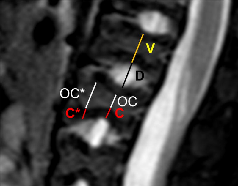
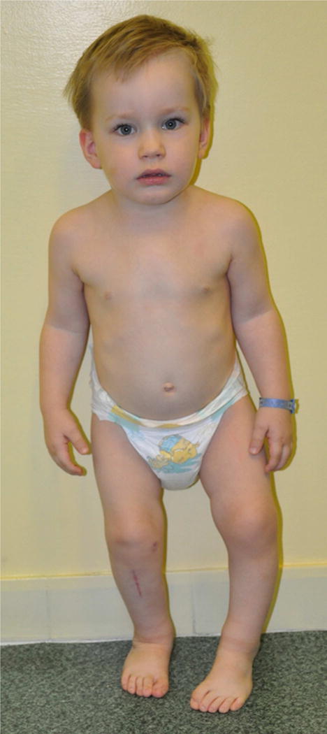
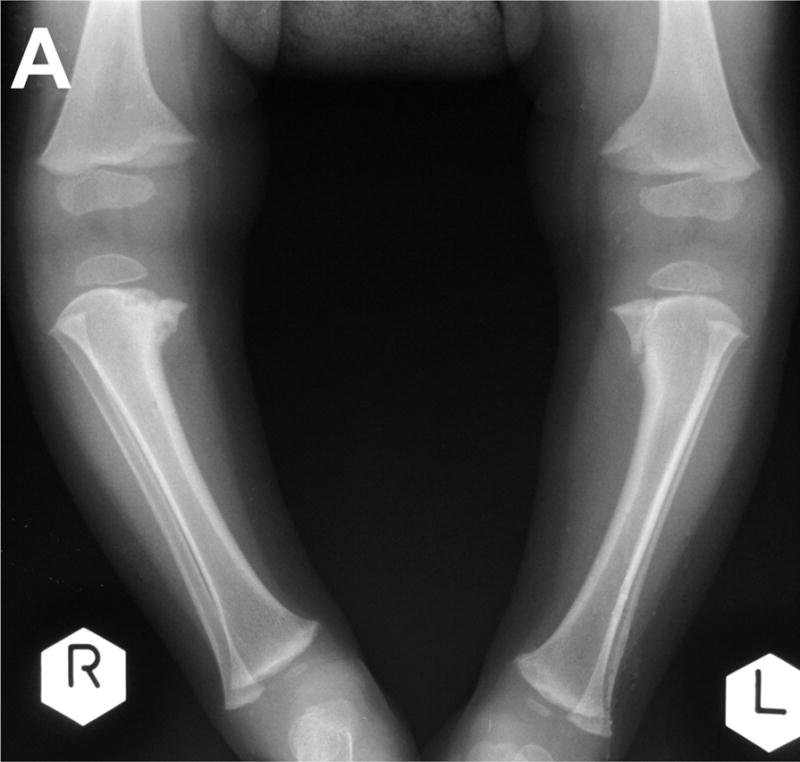
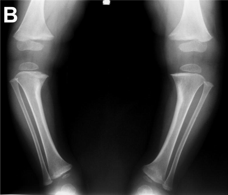
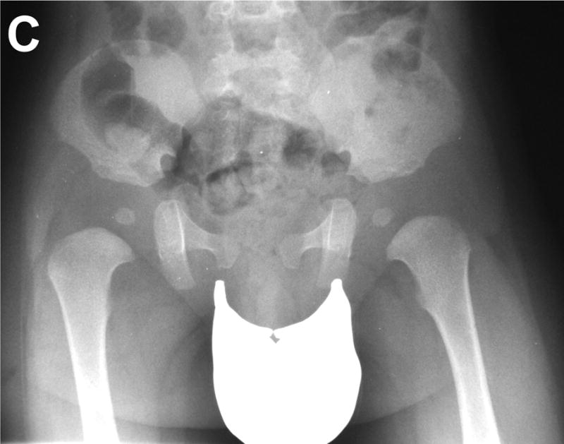
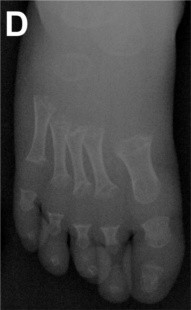
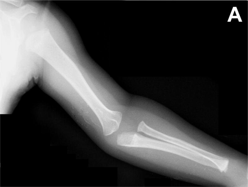
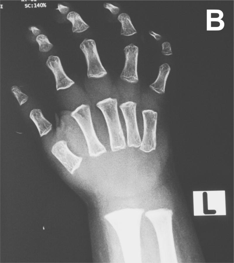
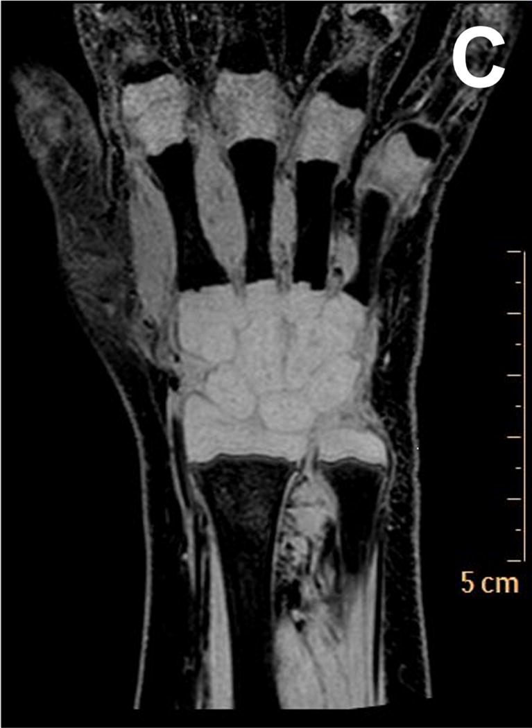
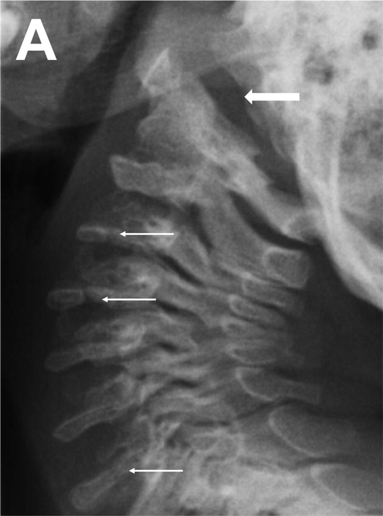
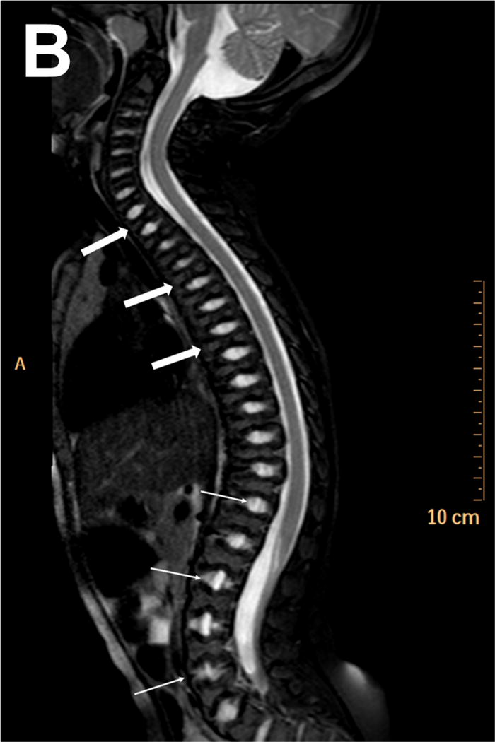
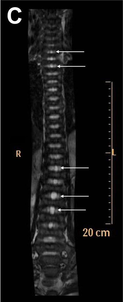
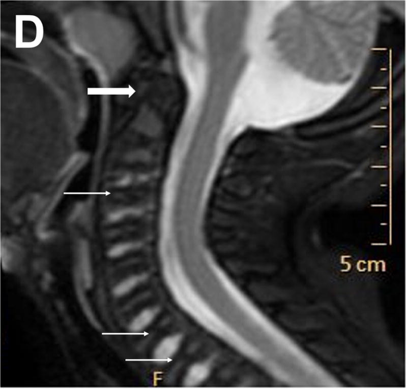
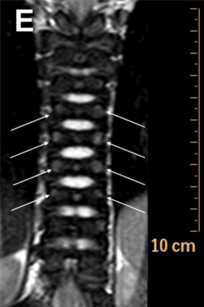
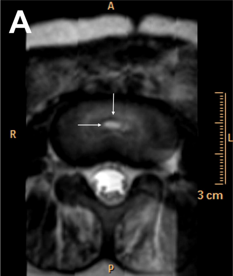
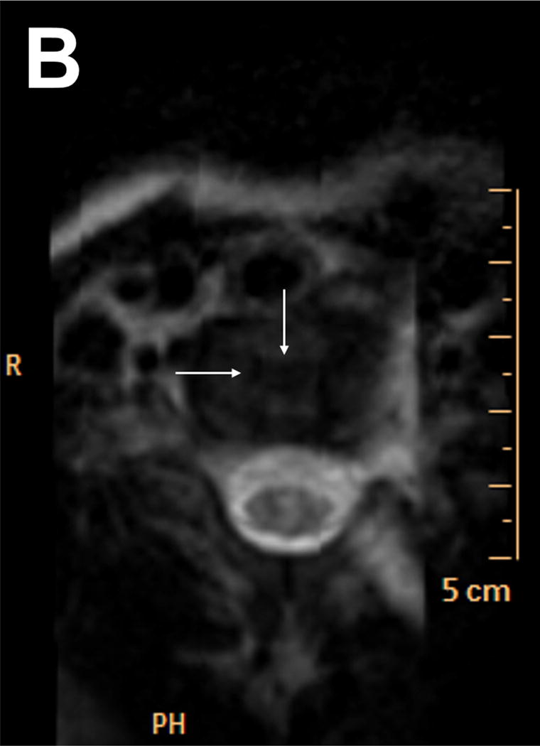
Spondylometaphyseal dysplasia Kozlowski type (SMDK) is a monogenic disorder within the TRPV4 dysplasia spectrum and has characteristic spinal and metaphyseal changes. We report skeletal MR imaging in a two-year-old patient who manifested typical clinical and radiographic features of SMDK. The diagnosis was confirmed by molecular analysis which revealed a mutation NM_021625.4:c.1781G > A - p.(Arg594His) in exon 11 of the TRPV4 gene. We have documented abnormalities in endochondral formation of the long and short tubular bones as well as round bones of the wrists and feet. The vertebral bodies had increased thickness of hyaline cartilage which enveloped ossification centers. The vertebrae and discs also had abnormalities in size, shape and structure. These anomalies were most likely the consequence of notochordal remnants presence within the intervertebral discs and in the vertebral bodies. The advantages of MR imaging in bone dysplasias caused by TRPV4 mutations are emphasized in this article.


Conflict of interest statement

CONFLICT OF INTERESTS

The authors declare that they have no competing financial interests.

Figures

Figure 1
Figure 1
Sagittal T2-weighted magnetic resonance image of the spine in midline demonstrating measurements of spinal structures used for ratios calculation: ossification center (OC), vertebral cartilage (C), disc (D), vertebral body (V) measurements obtained in the center of the vertebral bodies and intervertebral discs. *Measurements obtained at anterior 1/4 segment of vertebral bodies
Figure 2
Figure 2
Photograph of the patient at 26 months of age. Note shortening of the trunk, varus deformity of the left knee and the right tibia after surgical correction.
Figure 3
Figure 3
A: AP radiograph of bilateral lower extremities obtained at 22 months of age demonstrates bilateral genu varum with metaphyseal widening and fragmentation of the medial aspect of the proximal tibial metaphyses (corner pseudofractures). B: AP radiograph of bilateral knee taken at 25 months of age, demonstrates healing of the proximal medial tibial pseudofractures with improvement of the genu varum. C: AP pelvis radiograph obtained at age of 21 months shows: flared iliac wings, short sacroiliac notches, horizontal acetabula and short femoral necks with widening of the proximal femoral metaphyses and mild coxa vara. D: AP radiograph of the right foot at the age of three years demonstrates an apparent shortening of metatarsals with scalloping of metaphyses most pronounced distally. Broad first metatarsal and proximal phalanges; hypoplastic or absent middle and distal phalanges II – V.
Figure 3
Figure 3
A: AP radiograph of bilateral lower extremities obtained at 22 months of age demonstrates bilateral genu varum with metaphyseal widening and fragmentation of the medial aspect of the proximal tibial metaphyses (corner pseudofractures). B: AP radiograph of bilateral knee taken at 25 months of age, demonstrates healing of the proximal medial tibial pseudofractures with improvement of the genu varum. C: AP pelvis radiograph obtained at age of 21 months shows: flared iliac wings, short sacroiliac notches, horizontal acetabula and short femoral necks with widening of the proximal femoral metaphyses and mild coxa vara. D: AP radiograph of the right foot at the age of three years demonstrates an apparent shortening of metatarsals with scalloping of metaphyses most pronounced distally. Broad first metatarsal and proximal phalanges; hypoplastic or absent middle and distal phalanges II – V.
Figure 4
Figure 4
A: AP radiograph of the left upper extremity at the age 22 months shows broad metaphyses with undermodeling of the proximal shaft. B: AP radiograph of the left hand at the age 20 months with mild metaphyseal scalloping and delayed appearance of the carpal and epiphyseal ossification centers. C: MRI of the left hand at age 28 months. T1W image of WatSc sequence (TE 5.3 msec, TR 20 msec). Normal shape of the unossified carpal “bones” is observed.
Figure 4
Figure 4
A: AP radiograph of the left upper extremity at the age 22 months shows broad metaphyses with undermodeling of the proximal shaft. B: AP radiograph of the left hand at the age 20 months with mild metaphyseal scalloping and delayed appearance of the carpal and epiphyseal ossification centers. C: MRI of the left hand at age 28 months. T1W image of WatSc sequence (TE 5.3 msec, TR 20 msec). Normal shape of the unossified carpal “bones” is observed.
Figure 5
Figure 5
A: Lateral view of the cervical spine of three-year-old patient. Note platyspondyly with anteriorly rounded flat vertebral bodies and coronal clefts (thin arrows). Odontoid process is not visible giving the appearance of the odontoid hypoplasia (thick arrow). B–E: MRI of the entire spine in sagittal plane using using T2W SPAIR SENSE (TE 72 msec, TR 3536 msec) obtained at 28 months of age. B–D: Vertebral bodies are formed by ossification centers surrounded by vertebral hyaline cartilage. Thoracic and lumbar discs are concave with high signal intensity. Well-depicted high signal intensity structures are evident within the center of intervertebral discs representing notochord remnants (thin arrows). There are low signal intensity columns in the middle of the vertebrae thought to represent persistent notochord canals (thick arrows). C: The curve-plantar reformation was used for correction of the kyphoscoliosis and delineation of the entire vertebral column on a single coronal image. D: Sagittal view of the cervical spine with intravertebral, low signal intensity cartilaginous structures (thin arrows) corresponding to apparent vertebral body clefts demonstrated on the lateral radiographs (5A).The odontoid process is present but completely composed of cartilage therefore not visible on corresponding radiographs (thick arrow). E: Thoracic vertebral bodies with enlarged pedicles (arrows).
Figure 5
Figure 5
A: Lateral view of the cervical spine of three-year-old patient. Note platyspondyly with anteriorly rounded flat vertebral bodies and coronal clefts (thin arrows). Odontoid process is not visible giving the appearance of the odontoid hypoplasia (thick arrow). B–E: MRI of the entire spine in sagittal plane using using T2W SPAIR SENSE (TE 72 msec, TR 3536 msec) obtained at 28 months of age. B–D: Vertebral bodies are formed by ossification centers surrounded by vertebral hyaline cartilage. Thoracic and lumbar discs are concave with high signal intensity. Well-depicted high signal intensity structures are evident within the center of intervertebral discs representing notochord remnants (thin arrows). There are low signal intensity columns in the middle of the vertebrae thought to represent persistent notochord canals (thick arrows). C: The curve-plantar reformation was used for correction of the kyphoscoliosis and delineation of the entire vertebral column on a single coronal image. D: Sagittal view of the cervical spine with intravertebral, low signal intensity cartilaginous structures (thin arrows) corresponding to apparent vertebral body clefts demonstrated on the lateral radiographs (5A).The odontoid process is present but completely composed of cartilage therefore not visible on corresponding radiographs (thick arrow). E: Thoracic vertebral bodies with enlarged pedicles (arrows).
Figure 6
Figure 6
MR T2 weighted image of the lumbar spine in axial plane. A: An oval, high signal intensity structure in the center of the lumbar intervertebral disc thought to represent a notochordal remnant (arrows). B: There is a hypointensive structure within the lumbar vertebral body representing the notochordal canal (arrows).
Figure 6
Figure 6
MR T2 weighted image of the lumbar spine in axial plane. A: An oval, high signal intensity structure in the center of the lumbar intervertebral disc thought to represent a notochordal remnant (arrows). B: There is a hypointensive structure within the lumbar vertebral body representing the notochordal canal (arrows).

Similar articles

Cited by

References

    1. Andreucci E, Aftimos S, Alcausin M, Haan E, Hunter W, Kannu P, Kerr B, McGillivray G, Gardner RJM, Patricelli MG, Sillence D, Thompson E, Zacharin M, Zankl A, Lamandé SR, Savarirayan R. TRPV4 related skeletal dysplasias: a phenotypic spectrum highlighted by clinical, radiographic, and molecular studies in 21 new families. Orphanet J Rare Dis. 2011;6:37. - PMC - PubMed
    1. Camacho N, Krakow D, Johnykutty S, Katzman PJ, Pepkowitz S, Vriens J, Nilius B, Boyce BF, Cohn DH. Dominant TRPV4 mutations in nonlethal and lethal metatropic dysplasia. Am J Med Genet Part A. 2010;152A(5):1169–1177. - PMC - PubMed
    1. Cho TJ, Matsumoto K, Fano V, Dai J, Kim OH, Chae JH, Yoo WJ, Tanaka Y, Matsui Y, Takigami I, Monges S, Zabel B, Shimizu K, Nishimura G, Lausch E, Ikegawa S. TRPV4-pathy manifesting both skeletal dysplasia and peripheral neuropathy: a report of three patients. Am J Med Genet A. 2012;158A(4):795–802. - PubMed
    1. Christopherson LR, Rabin BM, Hallam DK, Russell EJ. Perisistence of the notochordal canal: MR and plain film appearance. AJNR Am J Neuroradiol. 1999;20:33–36. - PubMed
    1. Fleming A, Keynes RJ, Tannahill D. The role of notochord in vertebral column formation. J Anat. 2001;199:177–180. - PMC - PubMed

Publication types

MeSH terms

Supplementary concepts