. 2017 Nov;28(11):3191-3204.
doi: 10.1681/ASN.2016060690. Epub 2017 Jul 7.

Endothelial NF- κ B Blockade Abrogates ANCA-Induced GN

Affiliations

Endothelial NF- κ B Blockade Abrogates ANCA-Induced GN

Mira Choi et al. J Am Soc Nephrol. 2017 Nov.

Abstract

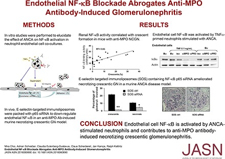
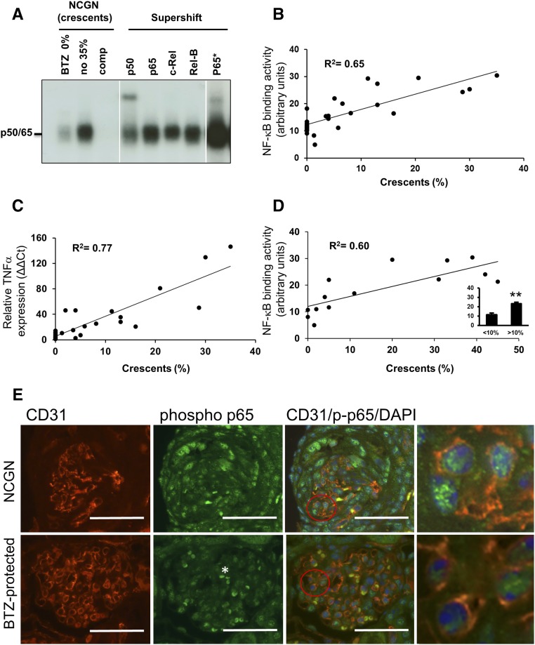
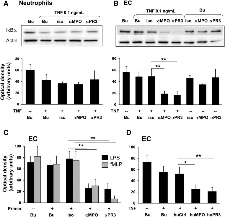
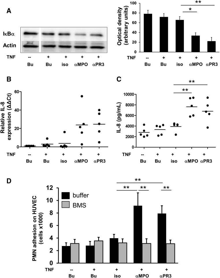
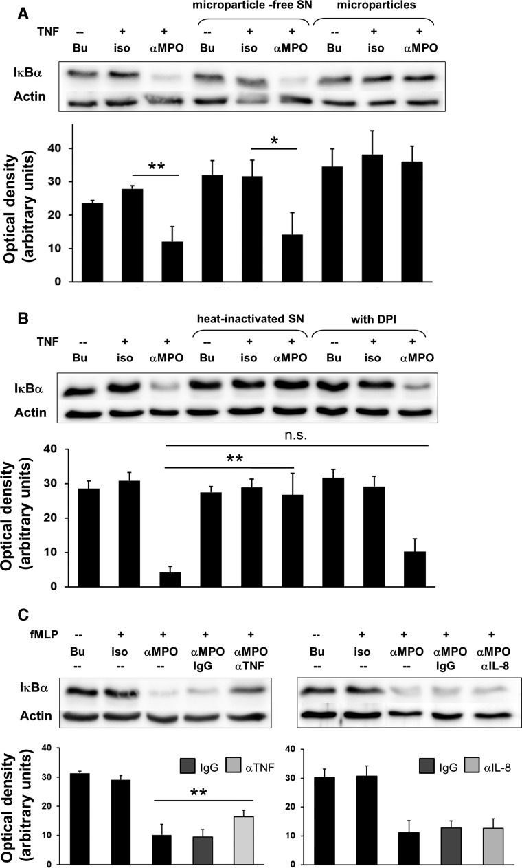
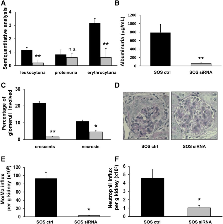
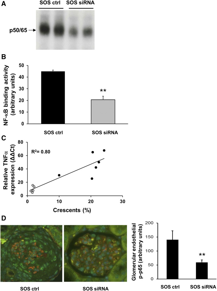
ANCA-associated vasculitis (AAV) is a highly inflammatory condition in which ANCA-activated neutrophils interact with the endothelium, resulting in necrotizing vasculitis. We tested the hypothesis that endothelial NF-κB mediates necrotizing crescentic GN (NCGN) and provides a specific treatment target. Reanalysis of kidneys from previously examined murine NCGN disease models revealed NF-κB activation in affected kidneys, mostly as a p50/p65 heterodimer, and increased renal expression of NF-κB-dependent tumor necrosis factor α (TNF-α). NF-κB activation positively correlated with crescent formation, and nuclear phospho-p65 staining showed NF-κB activation within CD31-expressing endothelial cells (ECs) in affected glomeruli. Therefore, we studied the effect of ANCA on NF-κB activation in neutrophil/EC cocultures in vitro ANCA did not activate NF-κB in primed human neutrophils, but ANCA-stimulated primed neutrophils activated NF-κB in ECs, at least in part via TNF-α release. This effect increased endothelial gene transcription and protein production of NF-κB-regulated interleukin-8. Moreover, upregulation of endothelial NF-κB promoted neutrophil adhesion to EC monolayers, an effect that was inhibited by a specific IKKβ inhibitor. In a murine NCGN model, prophylactic application of E-selectin-targeted immunoliposomes packed with p65 siRNA to downregulate endothelial NF-κB significantly reduced urine abnormalities, renal myeloid cell influx, and NCGN. Increased glomerular endothelial phospho-p65 staining in patients with AAV indicated that NF-κB is activated in human NCGN also. We suggest that ANCA-stimulated neutrophils activate endothelial NF-κB, which contributes to NCGN and provides a potential therapeutic target in AAV.

Keywords: ANCA; endothelium; glomerulonephritis; transcription factors; vasculitis.


Figures

None
Graphical abstract
Figure 1.
Figure 1.
NF-κB activity correlates with glomerular crescent formation in mice with anti-MPO antibody-induced NCGN. (A and B) Nuclear kidney extracts from untreated (n=6) or bortezomib (BTZ)-treated mice (n=22) in which we had induced NCGN using an anti-MPO antibody disease model were analyzed by EMSA. (A) Examples from a BTZ-treated mouse with 0% crescent formation (BTZ 0%) and an untreated mouse with 35% crescents (no 35%) are shown. Using the latter sample, we performed competition with a cold probe (comp) and supershift experiments using antibodies to p50, p65, c-Rel, and RelB as indicated. Note that longer exposure was needed to detect the p65 shift (p65*). (B) OD of the p50/p65 heterodimer was assessed in each sample using arbitrary units, and plotted against the percentage of glomerular crescents (n=28, correlation coefficient R2=0.65). (C) mRNA was prepared from the kidney of each mouse shown in (B), TNF-α mRNA expression was analyzed by quantitative RT-PCR and plotted against the percentage of glomerular crescents (n=28, correlation coefficient R2=0.77). (D) OD of the p50/p65 heterodimer was assessed in samples from NCGN mice that had not received active treatments and plotted against the percentage of glomerular crescents (n=14, correlation coefficient R2=0.60). The insert shows significantly less NF-κB activity in mice with <10% crescents (<10%) compared with those with >10% crescents (>10%). (E) Costaining using fluorescence-labeled antibodies to the EC marker CD31 (red) and phospho-p65 (green), together with DAPI staining of nuclei (blue) was performed in renal tissue from untreated mice with NCGN (NCGN) versus BTZ-protected mice (BTZ-protected). Weak phospho-p65 staining occurred in glomeruli from protected mice and strong signals in crescentic glomeruli of NCGN mice. Overlay images with magnifications in the very right panels indicate that some of the intense nuclear phospho-p65 staining in crescentic glomeruli was located within CD31-positive ECs. Low-powered images with scale bars, 50 μm; original magnification, ×40. A typical example is shown. **P<0.01.
Figure 2.
Figure 2.
Primed neutrophils stimulated with mAbs to MPO, PR3, and human ANCA IgG activate endothelial NF-κB in cocultures. A typical example for IκBα immunoblotting and the corresponding ODs (OD as arbitrary units) of the IκBα bands are provided for (A) lysates obtained from neutrophils stimulated with buffer control (Bu) or primed with 0.1 ng/ml TNF-α and subsequently treated for 60 minutes with Bu, an isotype control (iso), or mAbs to MPO (αMPO) and PR3 (αPR3), respectively (n=4); and (B) lysates obtained from ECs after 2 hours coculture with neutrophils that were treated with Bu or primed with 0.1 ng/ml TNF-α and subsequently incubated with Bu, iso, or αMPO and αPR3, respectively (n=8 for conditions shown in lanes 1–5, and n=4 for lanes 6–8). α-Actin served as a loading control. (C and D) OD measurements of the IκBα bands in lysates from ECs cocultured with neutrophils for 2 hours. (C) Neutrophils were either not primed or primed with 0.1 µg/ml LPS (black) or 10−8 M fMLP (gray) as indicated, subsequently incubated with αMPO and αPR3, respectively and added to the EC monolayers (n=6). (D) Neutrophils were either not primed or primed with TNF-α and subsequently incubated with Bu, human control IgG (huCtrl), MPO-ANCA (huMPO), or PR3-ANCA (huPR3), respectively, and added to the EC monolayers (n=5). *P<0.05; **P<0.01.
Figure 3.
Figure 3.
Endothelial NF-κB activation by ANCA-stimulated primed neutrophils does not require direct neutrophil/EC contact and leads to upregulation of IL-8, and increased neutrophil adhesion. (A–D) ECs were incubated with cellfree SN harvested from neutrophils treated for 5 hours with buffer (Bu) or primed with 0.1 ng/ml TNF-α and subsequently incubated with Bu, an isotype control (iso), or mAbs to MPO (αMPO) and PR3 (αPR3), respectively. All samples were analyzed on the same SDS gel and membrane. (A) ECs were incubated with cellfree neutrophil SN for 60 minutes and IκBα was assessed by immunoblotting. A typical example and the corresponding ODs (OD as arbitrary units) of the IκBα bands from all experiments are shown (n=5). α-Actin served as a loading control. (B) ECs were incubated with cellfree neutrophil SN for 6 hours and IL-8 mRNA was assessed by quantitative RT-PCR (n=5). (C) ECs were incubated with cellfree neutrophil SN for 18 hours and IL-8 protein was assessed in EC SN by ELISA (n=5). (D) ECs were preincubated with Bu (black) or the specific IKKβ inhibitor BMS (gray) for 60 minutes before cellfree neutrophil SN were added. After 18 hours, freshly isolated neutrophils were added, and neutrophil adhesion was determined after 120 minutes (n=5). *P<0.05; **P<0.01.
Figure 4.
Figure 4.
TNF-α released from ANCA-activated primed neutrophils contributes to endothelial NF-κB activation. (A) ECs were incubated for 60 minutes with cellfree SN harvested from neutrophils treated for 5 hours with buffer (Bu), or primed with 0.1 ng/ml TNF-α and subsequently incubated with Bu, an isotype control (iso), or mAb to MPO (αMPO), respectively (lanes 1–3). Microparticles were isolated from these SN, and either the microparticles or the microparticle-free SN were added to the ECs for 60 minutes (lane 4–9). IκBα was assessed by immunoblotting. A typical example and the corresponding ODs (OD as arbitrary units) of the IκBα bands from all experiments are shown (n=3). α-Actin served as a loading control. (B) Neutrophil SN preparation and EC treatment for samples in lane 1–3 was as described in (A). When indicated, SN were heat-inactivated or generated from 10 μM DPI-treated neutrophils (n=3). (C) Neutrophil SN preparation and EC treatment for samples in lane 1–3 were as described in (A). When indicated, SN were treated for 60 minutes with control IgG (IgG) or neutralizing antibodies for TNF-α (αTNF, left panel) or IL-8 (αIL-8, right panel). Note, fMLP was used as the priming agent in these experiments (n=4). *P<0.05; **P<0.01.
Figure 5.
Figure 5.
E-selectin targeted downregulation of the p65 NF-κB subunit using SOS p65 siRNA abrogates anti-MPO antibody-induced NCGN in mice. (A) Semiquantitative dipstick urine analysis from anti-MPO antibody-treated mice that received a control compound (black bars) or SOS p65 siRNA (gray bars). (B) Albuminuria was quantified by ELISA. (C) Histologic analysis of glomerular crescents formation and necrosis using periodic acid–Schiff staining, with a typical example given in (D), original magnification, ×40. (E and F) Flow cytometry of kidney tissue suspension to assess monocytes/macrophage and neutrophil influx into the kidneys of control-treated versus SOS p65 siRNA-treated mice. n=5 mice in each treatment group; *P<0.05; **P<0.01.
Figure 6.
Figure 6.
Preemptive SOS p65 siRNA treatment of mice inhibited glomerular NF-κB activation by EMSA and NF-κB–dependent TNF-α mRNA expression by quantitative RT-PCR that correlated with crescent formation. (A) Glomeruli were prepared from two control-treated (SOS ctrl) and two SOS p65 siRNA-treated (SOS siRNA) mice and NF-κB was assessed by EMSA. (B) Nuclear kidney extracts from SOS ctrl (black, n=5) and SOS siRNA (gray, n=5) mice were subjected to EMSA and the OD of the p50/65 heterodimer was assessed. (C) mRNA was prepared from the kidney of each mouse shown in (B), TNF-α mRNA expression was determined by qRT-PCR and plotted against the percentage of crescents. (D) Costaining using fluorescence-labeled antibodies to the EC marker CD31 (red), phospho-p65 (green), and DAPI staining for nuclei (blue) was performed in renal tissue from SOS ctrl– and SOS siRNA–treated mice. A typical microscopy example (original magnification, ×40) together with the corresponding quantitative assessment of nuclear phospho-p65 staining within CD31-positive glomerular ECs using Image J is depicted. **P<0.01.
Figure 7.
Figure 7.
NF-κB is activated in glomeruli of patients with AAV and active lesions. (A–D) Immunohistochemistry for phospho-p65 was performed in renal tissue from patients with tumor nephrectomy (labeled as normal tissue, n=5), ANCA-NCGN (n=5), anti-GBM NCGN (n=3), and hypertensive (n=3) and membranous nephropathy (n=2) as disease controls, respectively. (E) The corresponding quantifications are depicted. Glomeruli with active lesions (crescents/necrosis), normal appearance by light microscopy, and glomerular sclerosis were separately assessed in biopsies from patients with ANCA-NCGN. (F) Costaining using fluorescence-labeled antibodies to the EC marker CD31 (red), phospho-p65 (green), and DAPI staining for nuclei (blue) was performed in ANCA-GN biopsy samples (NCGN) and in normal tissue from tumor nephrectomies. A typical microscopy example (original magnification, ×40; scale bars = 50 μm) together with the corresponding quantitative assessment of nuclear phospho-p65 staining within CD31-positive glomerular ECs using Image J is depicted. **P<0.01.

Similar articles

Cited by

References

    1. Davies DJ, Moran JE, Niall JF, Ryan GB: Segmental necrotising glomerulonephritis with antineutrophil antibody: Possible arbovirus aetiology? Br Med J (Clin Res Ed) 285: 606, 1982 - PMC - PubMed
    1. Halbwachs L, Lesavre P: Endothelium-neutrophil interactions in ANCA-associated diseases. J Am Soc Nephrol 23: 1449–1461, 2012 - PMC - PubMed
    1. Radford DJ, Lord JM, Savage CO: The activation of the neutrophil respiratory burst by anti-neutrophil cytoplasm autoantibody (ANCA) from patients with systemic vasculitis requires tyrosine kinases and protein kinase C activation. Clin Exp Immunol 118: 171–179, 1999 - PMC - PubMed
    1. Kettritz R, Schreiber A, Luft FC, Haller H: Role of mitogen-activated protein kinases in activation of human neutrophils by antineutrophil cytoplasmic antibodies. J Am Soc Nephrol 12: 37–46, 2001 - PubMed
    1. Ben-Smith A, Dove SK, Martin A, Wakelam MJ, Savage CO: Antineutrophil cytoplasm autoantibodies from patients with systemic vasculitis activate neutrophils through distinct signaling cascades: Comparison with conventional Fcgamma receptor ligation. Blood 98: 1448–1455, 2001 - PubMed

MeSH terms

LinkOut - more resources