Meta-Analysis
. 2017 Aug 1;89(5):506-516.
doi: 10.1212/WNL.0000000000004176. Epub 2017 Jul 7.

A meta-analysis on progressive atrophy in intractable temporal lobe epilepsy: Time is brain?

Affiliations
Meta-Analysis

A meta-analysis on progressive atrophy in intractable temporal lobe epilepsy: Time is brain?

Lorenzo Caciagli et al. Neurology. .

Abstract

Objective: It remains unclear whether drug-resistant temporal lobe epilepsy (TLE) is associated with cumulative brain damage, with no expert consensus and no quantitative syntheses of the available evidence.

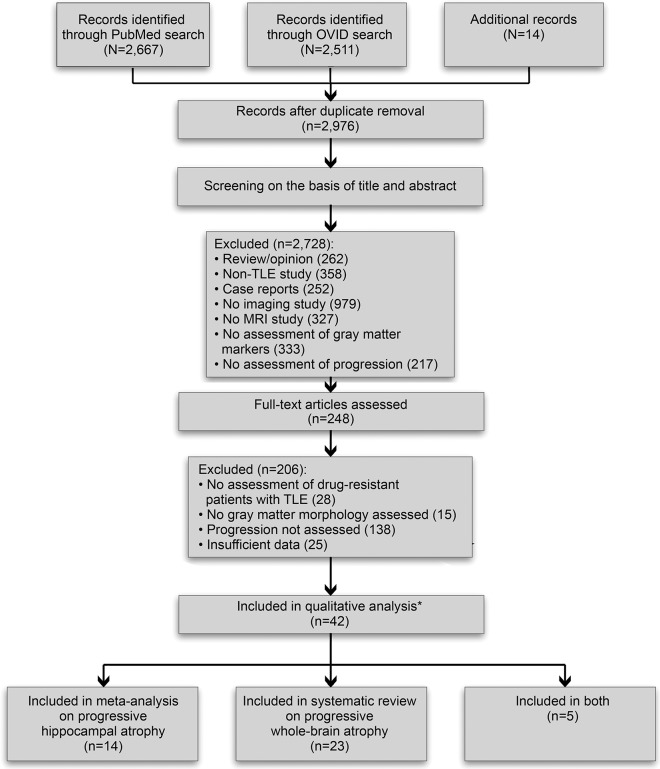
Methods: We conducted a systematic review and meta-analysis of MRI studies on progressive atrophy, searching PubMed and Ovid MEDLINE databases for cross-sectional and longitudinal quantitative MRI studies on drug-resistant TLE.

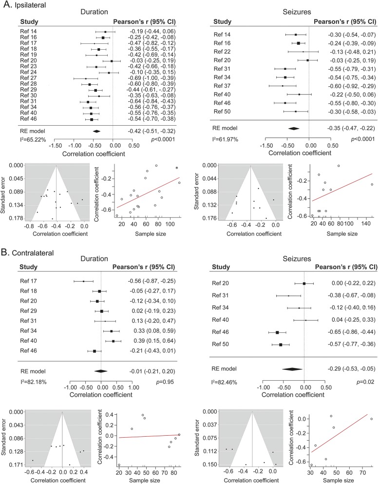
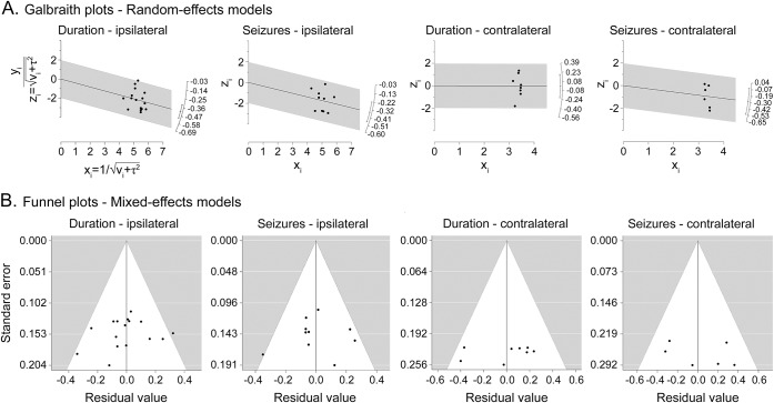
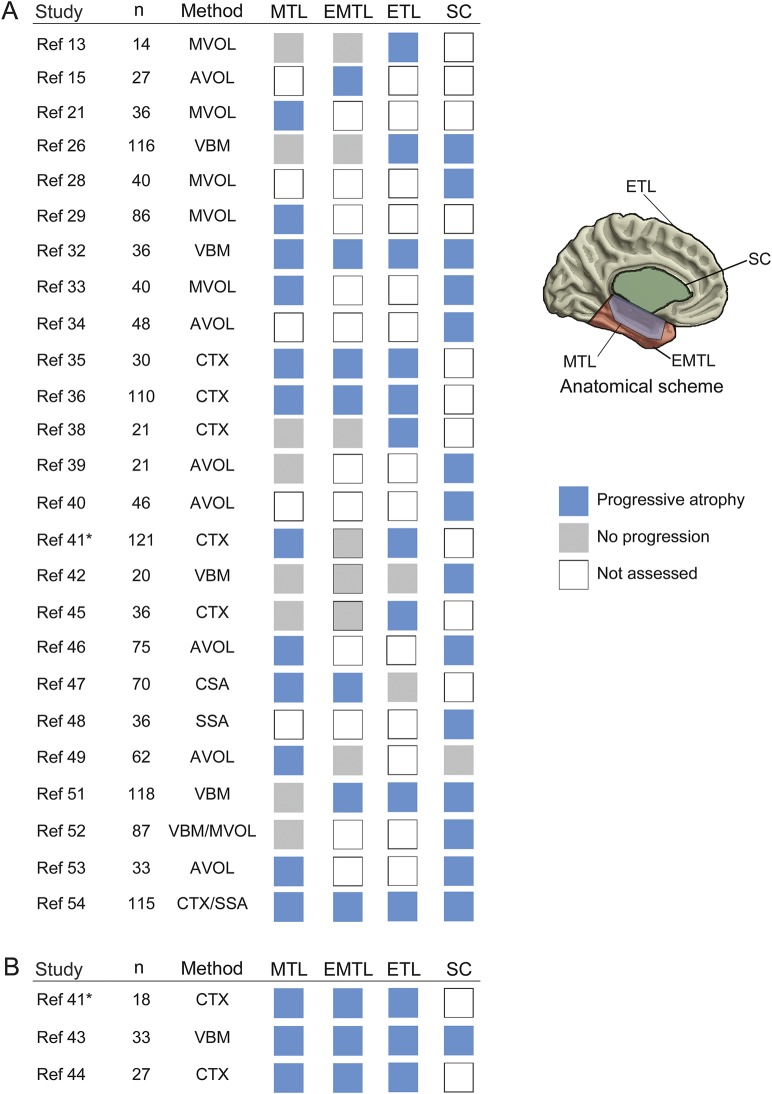
Results: We screened 2,976 records and assessed eligibility of 248 full-text articles. Forty-two articles met the inclusion criteria for quantitative evaluation. We observed a predominance of cross-sectional studies, use of different clinical indices of progression, and high heterogeneity in age-control procedures. Meta-analysis of 18/1 cross-sectional/longitudinal studies on hippocampal atrophy (n = 979 patients) yielded a pooled effect size of r = -0.42 for ipsilateral atrophy related to epilepsy duration (95% confidence interval [CI] -0.51 to -0.32; p < 0.0001; I2 = 65.22%) and r = -0.35 related to seizure frequency (95% CI -0.47 to -0.22; p < 0.0001; I2 = 61.97%). Sensitivity analyses did not change the results. Narrative synthesis of 25/3 cross-sectional/longitudinal studies on whole brain atrophy (n = 1,504 patients) indicated that >80% of articles reported duration-related progression in extratemporal cortical and subcortical regions. Detailed analysis of study design features yielded low to moderate levels of evidence for progressive atrophy across studies, mainly due to dominance of cross-sectional over longitudinal investigations, use of diverse measures of seizure estimates, and absence of consistent age control procedures.

Conclusions: While the neuroimaging literature is overall suggestive of progressive atrophy in drug-resistant TLE, published studies have employed rather weak designs to directly demonstrate it. Longitudinal multicohort studies are needed to unequivocally differentiate aging from disease progression.


Figures

Figure 1
Figure 1. Preferred Reporting Items for Systematic Reviews and Meta-Analyses flow diagram for the study screening and inclusion procedures
*References 16 and 25 refer to the same cohort, for which results are considered once only. Reference 41 contains separate cross-sectional and longitudinal analysis components, and is considered twice. TLE = temporal lobe epilepsy.
Figure 2
Figure 2. Meta-analysis on progressive hippocampal atrophy
Pooled effect size for the ipsilateral (A) and contralateral (B) hippocampus for studies that assessed the effects of epilepsy duration (left) and of seizure estimates (right) on hippocampal volumes. For each subsection, funnel plots (left side, relating study-wise effect size to standard error) and graphs for regression tests (right side, relating effect size to sample size) are provided. CI = confidence interval.
Figure 3
Figure 3. Galbraith (radial) plots for random effects models and funnel plots for mixed-effect models
(A) Galbraith (radial) plots for random effects models and (B) funnel plots for mixed-effect models. For mixed-effects models, study moderator is represented by volumetry technique (automated vs manual).
Figure 4
Figure 4. Systematic review on whole brain studies addressing progressive atrophy
Studies are divided into (A) cross-sectional and (B) longitudinal analyses. For each study, the sample size and quantitative MRI methods are provided as well as the reported finding (blue: progressive atrophy, gray: no progression, white: progression not assessed) across 4 brain subsystems (mesial temporal lobe [MTL], extramesial or lateral temporal lobe [EMTL], extratemporal cortical areas [ETL], subcortical gray matter [SC]). * Progressive mesio-temporal atrophy further documented at a subregional level in a subsequent analysis,e11 based on a largely overlapping cohort. For further information, see table e-1. AVOL = automated volumetry; CTX = cortical thickness analysis; MVOL = manual volumetry; SSA = surface-based shape analysis; VBM = voxel-based morphometry. McDonald 2008A and 2008B, Alhusaini 2012A and 2012B, and Keller 2015A and B refer to references 38, , , , , and , respectively.

References

    1. Wiebe S, Blume WT, Girvin JP, Eliasziw M; Effectiveness and Efficiency of Surgery for Temporal Lobe Epilepsy Study Group. A randomized, controlled trial of surgery for temporal-lobe epilepsy. N Engl J Med 2001;345:311–318. - PubMed
    1. Engel J Jr, McDermott MP, Wiebe S, et al. ; Early Randomized Surgical Epilepsy Trial (ERSET) Study Group. Early surgical therapy for drug-resistant temporal lobe epilepsy: a randomized trial. JAMA 2012;307:922–930. - PMC - PubMed
    1. Haneef Z, Stern J, Dewar S, Engel J Jr. Referral pattern for epilepsy surgery after evidence-based recommendations: a retrospective study. Neurology 2010;75:699–704. - PMC - PubMed
    1. Burneo JG, Shariff SZ, Liu K, Leonard S, Saposnik G, Garg AX. Disparities in surgery among patients with intractable epilepsy in a universal health system. Neurology 2016;86:72–78. - PMC - PubMed
    1. Wiebe S. Burden of intractable epilepsy. Adv Neurol 2006;97:1–4. - PubMed

MeSH terms