. 2017 Jul 7;7(1):4902.
doi: 10.1038/s41598-017-05315-y.

The interaction of the bioinsecticide PA1b (Pea Albumin 1 subunit b) with the insect V-ATPase triggers apoptosis

Affiliations

The interaction of the bioinsecticide PA1b (Pea Albumin 1 subunit b) with the insect V-ATPase triggers apoptosis

Vanessa Eyraud et al. Sci Rep. .

Abstract

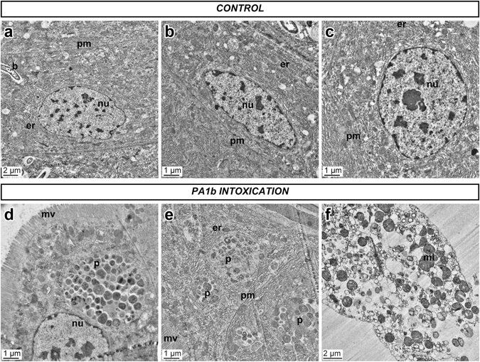
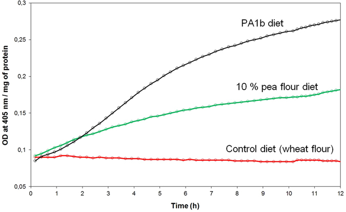
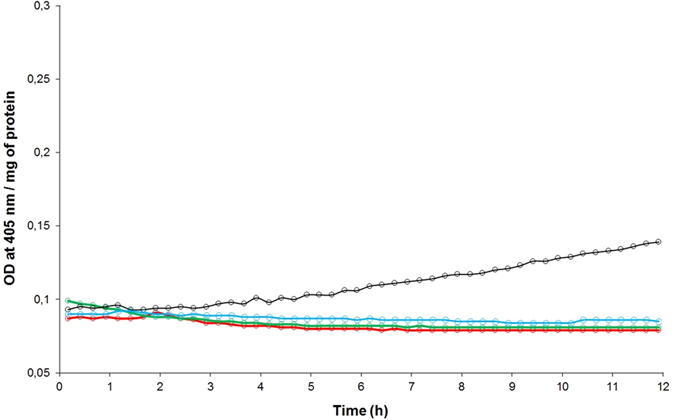
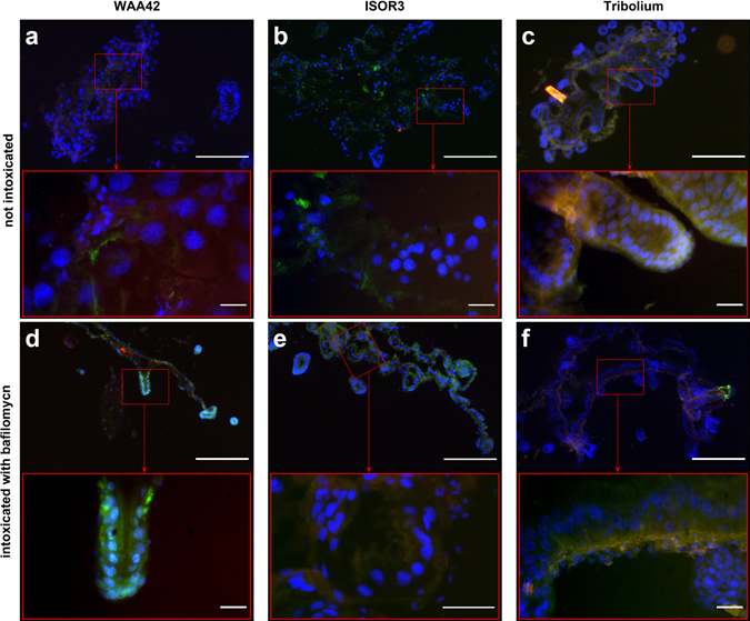
PA1b (Pea Albumin 1, subunit b) peptide is an entomotoxin, extracted from Legume seeds, with a lethal activity towards several insect pests, such as mosquitoes, some aphids and cereal weevils. This toxin acts by binding to the subunits c and e of the plasma membrane H+-ATPase (V-ATPase) in the insect midgut. In this study, two cereal weevils, the sensitive Sitophilus oryzae strain WAA42, the resistance Sitophilus oryzae strain ISOR3 and the insensitive red flour beetle Tribolium castaneum, were used in biochemical and histological experiments to demonstrate that a PA1b/V-ATPase interaction triggers the apoptosis mechanism, resulting in insect death. Upon intoxication with PA1b, apoptotic bodies are formed in the cells of the insect midgut. In addition, caspase-3 enzyme activity occurs in the midgut of sensitive weevils after intoxication with active PA1b, but not in the midgut of resistant weevils. These biochemical data were confirmed by immuno-histochemical detection of the caspase-3 active form in the midgut of sensitive weevils. Immuno-labelling experiments also revealed that the caspase-3 active form and V-ATPase are close-localized in the insect midgut. The results concerning this unique peptidic V-ATPase inhibitor pave the way for the utilization of PA1b as a promising, more selective and eco-friendly insecticide.


Conflict of interest statement

The authors declare that they have no competing interests.

Figures

Figure 1
Figure 1
Midgut cells from a PA1b sensitive S. oryzae strain (WAA42) observed with transmission electron microscopy. Insect midguts were dissected, fixed, and ultra-thin sections (70 nm) were prepared as described in the Experimental Procedures section. The sensitive weevil WAA42 was fed on wheat flour either without (Control, upper panel a,b,c), or with PA1b (400 µg per g of food) for 24 h (lower panel, d,e,f). b: bacterium; er: endoplasmic reticulum; mi: mitochondria; mv: microvilli; nu: nuclei; p: phagosome; pm: plasma membrane.
Figure 2
Figure 2
Caspase-3 activity on weevil midguts following PA1b intoxication. The weevils of the PA1b sensitive strain WAA42 were intoxicated for 24 h with an artificial diet composed of wheat flour (control, red curve); PA1b incorporated in the pea flour (10%, green curve); or PA1b (400 µg/g of food, black curve). After intoxication, the midguts were dissected and the caspase -3 activities were measured using the artificial substrate DEVD-pNA.
Figure 3
Figure 3
Induction kinetics of the caspase-3 activity by PA1b. The weevils of the PA1b sensitive strain WAA42 were intoxicated for different time periods (0, 3, 6, 12, 24, 48, 72 and 96 h) with an artificial diet composed of wheat flour incorporating PA1b (400 µg/g of food). After intoxication, the midguts were dissected and the caspase -3 activities were measured using the artificial substrate DEVD-pNA.
Figure 4
Figure 4
Specificity of the caspase-3 activity induced by PA1b. The weevils were intoxicated for 24 h with PA1b, 400 µg per g of food. After intoxication, the midguts were dissected and the caspase -3 activities were measured using the artificial substrate DEVD-pNA. (a) Sensitive weevil strain WAA42 intoxicated with PA1b (black) and control (without toxin, red); resistant weevil strain ISOR3 intoxicated with PA1b (blue) and control (green). (b) Sensitive weevil strain WAA42 intoxicated with PA1b (black); with reduced and alkylated PA1b (green); with the inactive mutant F10A (blue); and control (red). (c) Sensitive weevil strain WAA42 intoxicated with PA1b (black); control (red); with bafilomycin at 0.25 mM (dark blue) and at 0.8 mM (light blue); resistant weevil strain ISOR3 intoxicated with bafilomycin at 0.8 mM (green).
Figure 5
Figure 5
Caspase-3 activity on T. castaneum midguts following PA1b intoxication. The red flour beetles were intoxicated for 24 h with an artificial diet composed of 95% wheat flour and 5% yeast extract (control, blue curve). PA1b was incorporated in the diet (400 µg per g, green curve); or pea flour containing PA1b was added to the diet (10%, red curve or 25% black curve). After intoxication, the midguts were dissected and the caspase-3 activities were measured using the artificial substrate DEVD-pNA.
Figure 6
Figure 6
Immuno-localization of the caspase-3 active form in sensitive S. oryzae (strain WAA42) intoxicated by PA1b. Midguts of the sensitive weevil WAA42 were dissected, embedded, frozen and then tissue sections (7 µm) were cut. Slides were prepared as described in the Experimental Procedures section. Caspase-3 was labelled with rabbit anti-cleaved caspase-3 and revealed using a secondary antibody Alexa fluor 488 donkey anti-rabbit IgG. DAPI was added for nuclear staining. (a) Control (no PA1b); (b) weevils intoxicated with PA1b, at 400 µg per g of food, for 4 days; (c) weevils intoxicated with the inactive mutant of PA1b, R21A, at 400 µg per g of food, for 4 days. Upper panel scale bar = 200 µm; lower panel scale bar = 20 µm. Blue: nuclei stained with DAPI; green: caspase 3; brownish-red: non-specific signal from background autofluorescence. mc: mesenteric caeca.
Figure 7
Figure 7
Immuno-localization of the caspase-3 active form in resistant S. oryzae (strain ISOR3) and in the insensitive insect T. castaneum, intoxicated with PA1b. Insect midguts were dissected, embedded, frozen and then tissue sections (7 µm) were cut. Slides were prepared as described in the Experimental Procedures section. Caspase-3 was labelled with rabbit anti-cleaved caspase-3 and revealed using a secondary antibody Alexa fluor 488 donkey anti-rabbit IgG. DAPI was added for nuclear staining. The midgut of the resistant weevil strain ISOR3 reared on wheat flour (control, a) or intoxicated with PA1b, at 400 µg per g of food, for 4 days (b); the midgut of the insensitive insect T. castaneum reared on wheat flour (control, c) or intoxicated with PA1b, at 400 µg per g of food, for 4 days (d). Upper panel scale bar = 200 µm; lower panel scale bar = 20 µm. Blue: nuclei stained with DAPI; green: caspase 3; brownish-red: non-specific signal from background autofluorescence.
Figure 8
Figure 8
Immuno-localization of the caspase-3 active form and V-ATPase in sensitive S. oryzae (strain WAA42) intoxicated with PA1b. Midguts of the sensitive weevil WAA42 were dissected, embedded, frozen and then tissue sections (7 µm) were cut. Slides were prepared as described in the Experimental Procedures section. Caspase-3 was labelled with rabbit anti-cleaved caspase-3 and V-ATPase was labelled with a rabbit anti-ductin antibody. Revelation of both antibodies was performed using a secondary antibody Alexa fluor 488 donkey anti-rabbit IgG. To compare the labelling of caspase-3 and V-ATPase, labelling was performed on successive cuts and the green signal of anti-V-ATPase was transformed into a red signal. DAPI was added for nuclear staining. (a) negative control (non-immune serum); (b) anti-V-ATPase antibody labelling and (c) anti-caspase-3 antibody labelling. Upper panel scale bar = 200 µm; lower panel scale bar = 20 µm. Blue: nuclei stained with DAPI; green: caspase 3; red: V-ATPase.
Figure 9
Figure 9
Immuno-localization of the caspase-3 active form in sensitive S. oryzae (strain WAA42), in resistant S. oryzae (ISOR3), and in insensitive T. castaneum intoxicated with bafilomycin. Midguts of insects were dissected, embedded, frozen and then tissue sections (7 µm) were cut. Slides were prepared as described in the Experimental Procedures section. Caspase-3 was labelled with rabbit anti-cleaved caspase-3, and revelation was performed using a secondary antibody Alexa fluor 488 donkey anti-rabbit IgG. DAPI was added for nuclear staining. The S. oryzae PA1b sensitive strain (WAA42), the resistant (ISOR3) strain, and the insensitive insect T. castaneum were all reared on non-intoxicated medium (panel a,b and c, respectively) or on the same medium containing bafilomycin (150 µg per g of food) (panel d, e, and f, respectively). Upper panel scale bar = 200 µm; lower panel scale bar = 20 µm. Blue: nuclei stained with DAPI; green: caspase 3; brownish-red: non-specific signal from background autofluorescence.

Similar articles

Cited by

References

    1. Becker-Ritt AB, Carlini CR. Fungitoxic and insecticidal plant polypeptides. Biopolymers. 2012;98:367–384. doi: 10.1002/bip.22097. - DOI - PubMed
    1. Colovic M, et al. Toxic effects of diazinon and its photodegradation products. Toxicology letters. 2010;193:9–18. doi: 10.1016/j.toxlet.2009.11.022. - DOI - PubMed
    1. Delobel, B., Grenier, A. M., Gueguen, J., Ferrasson, E. & Mbaiguinam, M. Utilisation d’un polypeptide dérivé d’une albumine PA1b de légumineuse comme insecticide (Brevet-98/05877). Paris patent (1998).
    1. Gressent, F., Da Silva, P., Eyraud, V., Karaki, L. & Royer, C. Pea Albumin 1 subunit b (PA1b), a promising bioinsecticide of plant origin. Toxins (Basel)3, 1502–1517, doi:10.3390/toxins3121502 (2011). - PMC - PubMed
    1. Gressent F, et al. Biological activity and binding site characteristics of the PA1b Entomotoxin on insects from different orders. J Insect Sci. 2007;7:1–10. doi: 10.1673/031.007.1201. - DOI - PMC - PubMed

MeSH terms