. 2017 Jul 7;7(1):4860.
doi: 10.1038/s41598-017-04071-3.

The Anti-Hiv Candidate Abx464 Dampens Intestinal Inflammation by Triggering Il-22 Production in Activated Macrophages

Affiliations

The Anti-Hiv Candidate Abx464 Dampens Intestinal Inflammation by Triggering Il-22 Production in Activated Macrophages

Karim Chebli et al. Sci Rep. .

Abstract

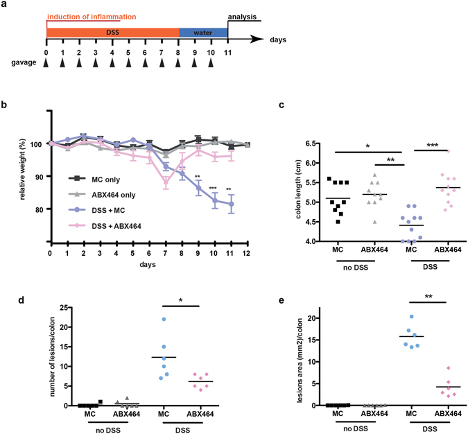
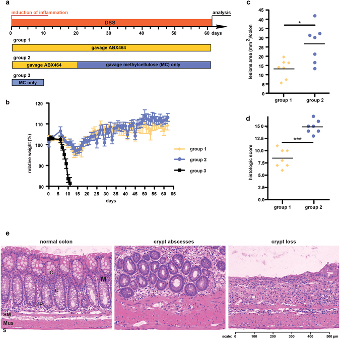
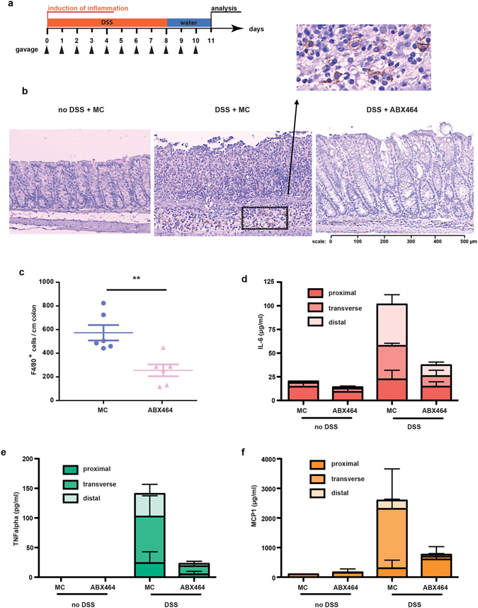
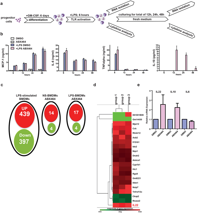
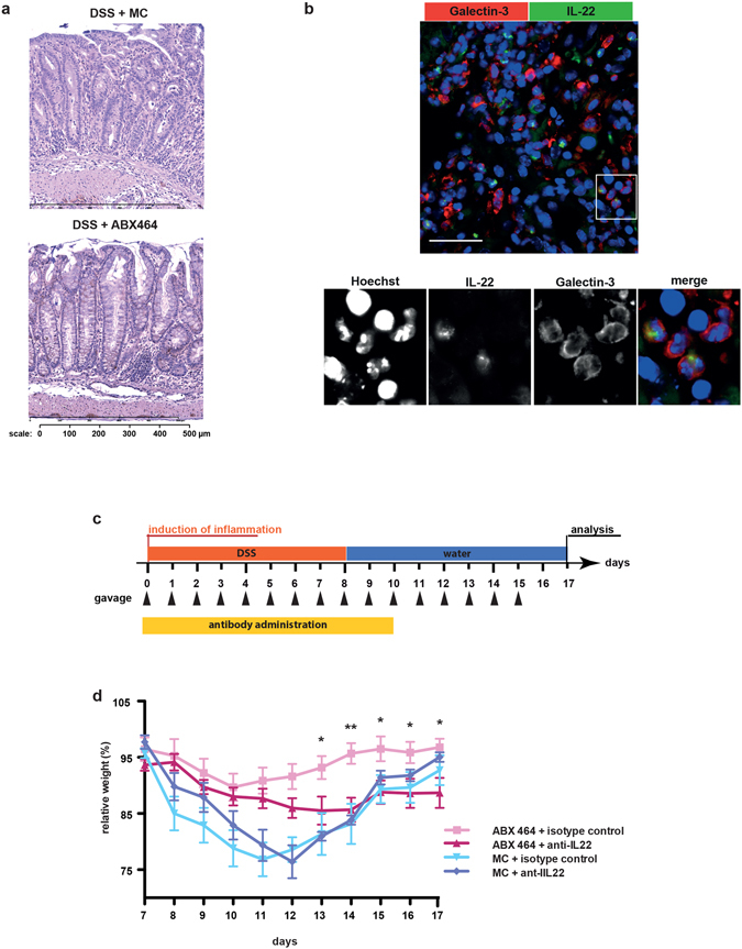
The progression of human immunodeficiency virus (HIV) is associated with mucosal damage in the gastrointestinal (GI) tract. This damage enables bacterial translocation from the gut and leads to subsequent inflammation. Dextran sulfate sodium (DSS-exposure) is an established animal model for experimental colitis that was recently shown to recapitulate the link between GI-tract damage and pathogenic features of SIV infection. The current study tested the protective properties of ABX464, a first-in-class anti-HIV drug candidate currently in phase II clinical trials. ABX464 treatment strongly attenuated DSS-induced colitis in mice and produced a long-term protection against prolonged DSS-exposure after drug cessation. Consistently, ABX464 reduced the colonic production of the inflammatory cytokines IL-6 and TNFα as well as that of the chemoattractant MCP-1. However, RNA profiling analysis revealed the capacity of ABX464 to induce the expression of IL-22, a cytokine involved in colitis tissue repair, both in DSS-treated mice and in LPS-stimulated bone marrow-derived macrophages. Importantly, anti-IL-22 antibodies significantly reduced the protective effect of ABX464 on colitis in DSS-treated mice. Because reduced IL-22 production in the gut mucosa is an established factor of HIV and DSS-induced immunopathogenesis, our data suggest that the anti-inflammatory properties of ABX464 warrant exploration in both HIV and inflammatory ulcerative colitis (UC) disease.


Conflict of interest statement

D.S., A.G., N.C. and H.J.E. are Abivax employees and hold Abivax stock options. J.T. is a member of the ABIVAX scientific advisory board and the collaborative laboratory that has received financial support from Abivax.

Figures

Figure 1
Figure 1
ABX464 treatment suppresses disease severity in DSS-induced colitis. (a) C57BL/6 mice (n = 12 each cohort) subjected to the DSS colitis protocol shown received orally once a day ABX464 (50 mg/kg) in methylcellulose or methylcellulose only through gavage. (b) Weight development in ABX464 and methycellulose (MC) only treated mice during DSS-induced colitis. Control cohorts included mice not exposed to DSS. (ce) At the end of the protocol described in (a) mice were sacrificed and colons were analyzed by histology for colitis severity including number of lesion (c), lesion size (d) and colon size (e).
Figure 2
Figure 2
The protective effect of ABX464 is maintained in mice during continuous DSS-exposure. (a) C57BL/6 mice (n = 8 each cohort) were challenged with DSS for 63 days. Mice received orally once a day ABX464 in methylcellulose during the whole protocol (group 1) or only for the first 20 days followed by methylcellulose only administration (group 2). Mice of group 3 received methylcellulose during DSS-exposure and had to be scarified at day 10. (b) Weight development in ABX464-treated mice during DSS-administration. (c,d) Colons were analyzed by histology for lesion size (c) and colitis severity (d) at the end of the protocol. Histological scoring was performed as described in the Material and Methods section and detailed in Supplementary Table 1. (e) Representative images for normal colon, crypt abscesses and crypt loss, the latter two being part of the histological scoring shown in 2d. Normal histology of the colon (normal colon); Mucosa, M, composed of epithelial crypt cells, C, underlined by lamina propria, LP; Submucosae, SM; Musculae, Mus; Serosa, S.
Figure 3
Figure 3
ABX464 decreases macrophage presence and pro-inflammatory cytokine production during acute colitis. (a) Acute colitis was induced as described in Fig. 1a. (b) Colons were analyzed for macrophage content by F4/80 labeling. Representative images of macrophage presence in colons of MC only treated mice (left hand panel), MC and ABX464 treated mice exposed to DSS are presented. (c) Total number of F4/80 + macrophages per colon per mouse are are shown (n = 8 each cohort). (d–f) Colons were taken at the end of the protocol, dissected in the proximal, middle and distal region and maintained in culture for 24 hours. Culture supernatant were analyzed for production of IL-6 (d), TNFalpha (e), and MCP-1 (f) by CBA as described in Methods section.
Figure 4
Figure 4
ABX464 treatment restores the expression of cytokine and chemokine signature in the colon of DSS-treated mice. (a) Identification of cytokines and chemokines regulated by ABX464 in colons of DSS-treated mice. RNA samples of from colons were taken at day 11 from mice exposed to DSS-exposure according to the protocol shown in Fig. 1a. Heatmaps show differential gene expression between colons of methylcellulose- and ABX464-treated mice as monitored by RT2 profiler PCR array (Qiagen). Scatterblots correlate gene expression in colons of ABX464-treated mice (a), methylcellulose (MC)- and DSS-exposed mice (b) and ABX464- and DSS-exposed mice (c) versus MC only treated mice. Samples represent the pool of RNA samples of n = 6 for each mouse cohort. (d) The clustergram shows differential gene expression between colons of MC- and ABX464-treated mice as monitored by RT2 profiler PCR array (Qiagen). Controls (Co) are tissue samples from mice not treated with DSS. Arrow heads indicate expression level of IL-22.
Figure 5
Figure 5
Effect of ABX464 on cytokine secretion and mRNA expression in BMDMs (a) Bone marrow isolated cells were cultured for 6 days in the presence of GM-CSF (50ng/ml) to differentiate into macrophages. Cells were kept in culture for additional 3 days in the presence of ABX464 (5 µM) or vehicle (DMSO) alone and for additional 6 hours stimulated with LPS (4 µg/ml). Cells were then kept in normal medium for additional 42 hours. Cell aliquots for RNA isolation and supernatants were taken at time 6, 12, 24 and 48 hours. (b) Culture supernatants of the indicated cell cultures were analyzed by CBA for the content of MCP-1, IL-6, TNFalpha and IL-10 as described in Materials and Methods. (c) A Venn diagram displaying the number of inducible or repressible (≥1.5 log2-fold) genes after 6 hours stimulation of LPS (LPS-stimulated BMDMs), non-stimulated BMDMs treated with ABX464 (NS-BMDMs ABX464) or LPS-stimulated BMDMs treated with ABX464 (LPS-BMDMs ABX464). (d) Identification of genes regulated by ABX464 in LPS-stimulated BMDMs. RNA samples of three group of cells taken at time point 12 h were analyzed by RNAseq and each group represents the relative expression between ABX464 and control (DMSO) treated samples. Only those genes are represented that were differentially expressed in each group. (e) Validation of elevated transcript levels of IL-22 in ABX464-treated LPS-stimulated BMDMs by qPCR.
Figure 6
Figure 6
The colitis dampening effect of ABX464 is partially mediated by IL-22 (a) Elevated protein levels of IL-22 are detectable on paraffin sections of colons taken from ABX464 treated mice. Colons were isolated at day 11 from mice exposed to DSS-exposure according to the protocol shown in Fig. 1a. (b) Confocal images of paraffin-sections from a colon of an ABX464-treated mouse as described in Fig. 6a. Section was stained for galactin-3 to identify macrophages (red) and IL-22 (green). Nuclei were stained with Hoechst (blue). The selected images shown are representative. Scale bars represent 100 µm the panel show 4x blow-ups. The cells shown in the blow up are located in the lamina propria between perturbed crypts. (c) C57BL/6 mice (n = 7 each cohort) were subjected to the DSS colitis protocol shown. received orally once a day ABX464 in methylcellulose or methylcellulose (MC) only through gavage. ABX464 and MC-treated were divided into two groups receiving daily 100 µg of either antagonistic anti-IL-22 antibody or isotype control. (d) Weight development in ABX464 and methycellulose (MC) only treated mice during DSS-induced colitis in the presence of either control or anti-IL-22 antibody.

References

    1. Scherrer, D. et al. Randomized Trial of Food Effect on Pharmacokinetic Parameters of ABX464 Administered Orally to Healthy Male Subjects Antimicrob. Agents Chemother. 61, e01288–16 (2017). - PMC - PubMed
    1. Scherrer, D. et al. Pharmacokinetics and tolerability of ABX464, a novel first in class compound to treat HIV infection, in healthy HIV- uninfected subjects. Journal of Antimicrobial Chemotherapy72, 820–828 (2017). - PubMed
    1. Campos N, et al. Long lasting control of viral rebound with a new drug ABX464 targeting Rev - mediated viral RNA biogenesis. Retrovirology. 2015;12:30. doi: 10.1186/s12977-015-0159-3. - DOI - PMC - PubMed
    1. Deeks SG, Tracy R, Douek DC. Systemic effects of inflammation on health during chronic HIV infection. Immunity. 2013;39:633–645. doi: 10.1016/j.immuni.2013.10.001. - DOI - PMC - PubMed
    1. Utay NS, Hunt PW. Role of immune activation in progression to AIDS. Curr Opin HIV AIDS. 2016;11:131–137. doi: 10.1097/COH.0000000000000242. - DOI - PMC - PubMed

Publication types

MeSH terms