. 2017 Jul 7;7(1):4916.
doi: 10.1038/s41598-017-04982-1.

Efficient activation of the lymphangiogenic growth factor VEGF-C requires the C-terminal domain of VEGF-C and the N-terminal domain of CCBE1

Affiliations

Efficient activation of the lymphangiogenic growth factor VEGF-C requires the C-terminal domain of VEGF-C and the N-terminal domain of CCBE1

Sawan Kumar Jha et al. Sci Rep. .

Abstract

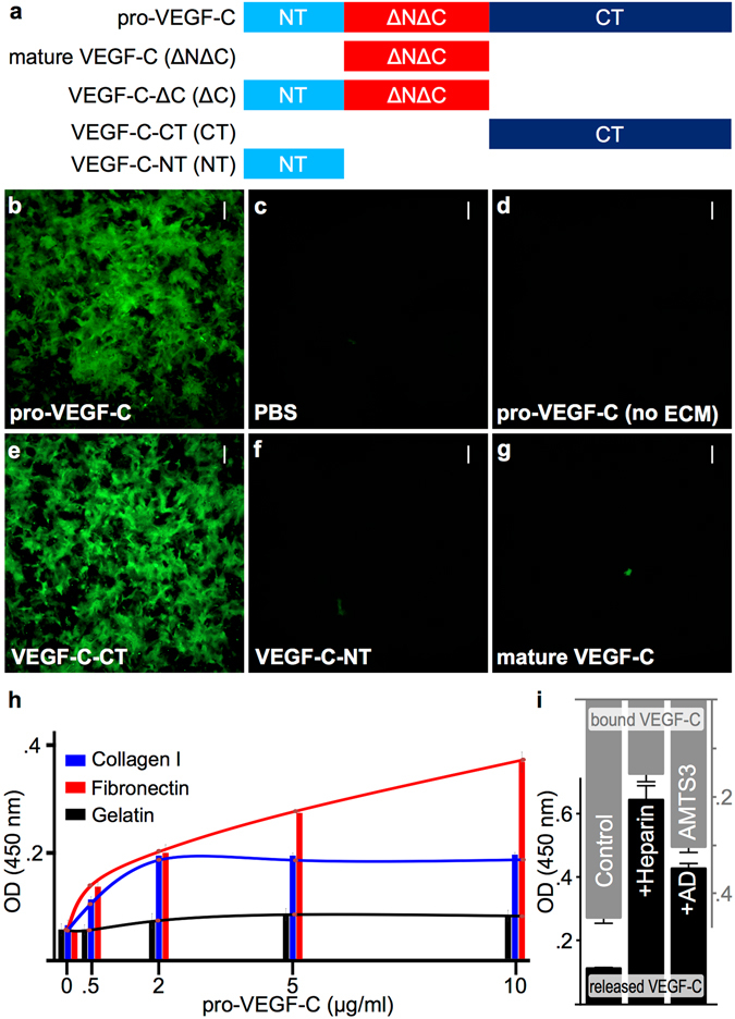
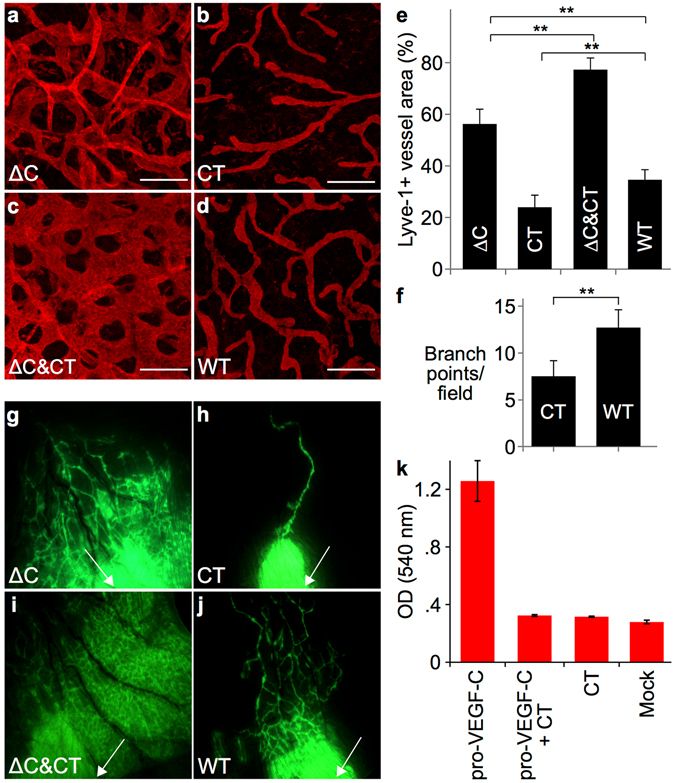
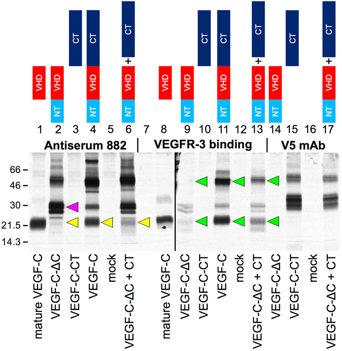
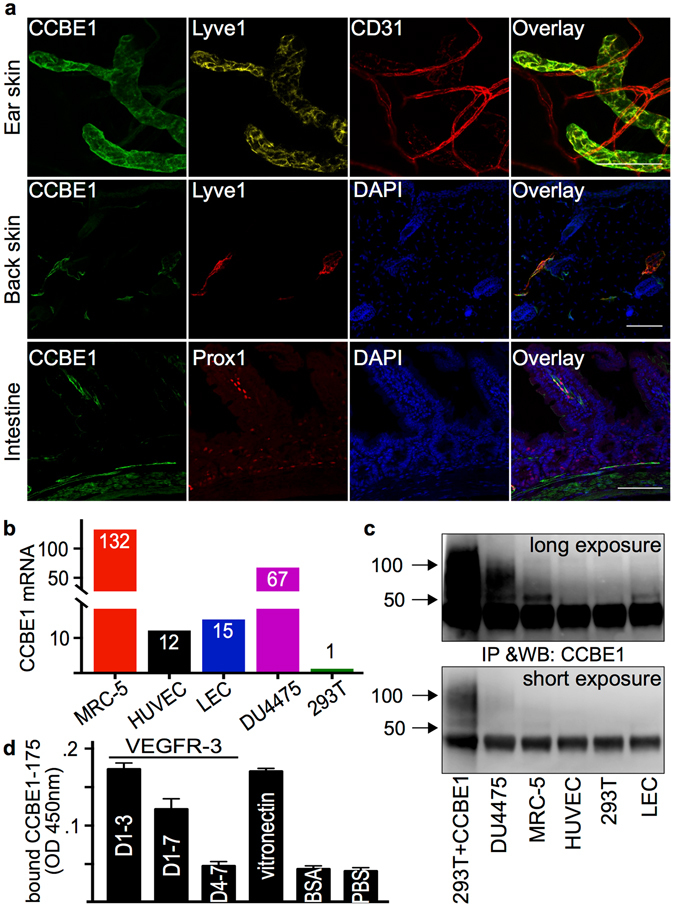
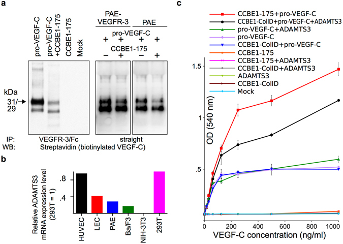
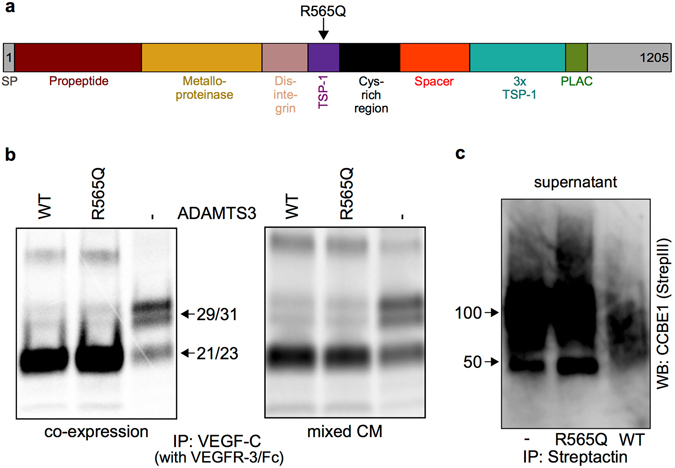
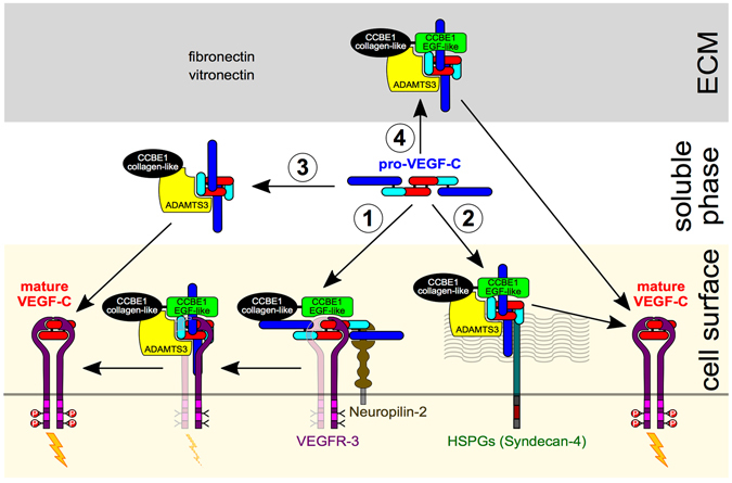
The collagen- and calcium-binding EGF domains 1 (CCBE1) protein is necessary for lymphangiogenesis. Its C-terminal collagen-like domain was shown to be required for the activation of the major lymphangiogenic growth factor VEGF-C (Vascular Endothelial Growth Factor-C) along with the ADAMTS3 (A Disintegrin And Metalloproteinase with Thrombospondin Motifs-3) protease. However, it remained unclear how the N-terminal domain of CCBE1 contributed to lymphangiogenic signaling. Here, we show that efficient activation of VEGF-C requires its C-terminal domain both in vitro and in a transgenic mouse model. The N-terminal EGF-like domain of CCBE1 increased VEGFR-3 signaling by colocalizing pro-VEGF-C with its activating protease to the lymphatic endothelial cell surface. When the ADAMTS3 amounts were limited, proteolytic activation of pro-VEGF-C was supported by the N-terminal domain of CCBE1, but not by its C-terminal domain. A single amino acid substitution in ADAMTS3, identified from a lymphedema patient, was associated with abnormal CCBE1 localization. These results show that CCBE1 promotes VEGFR-3 signaling and lymphangiogenesis by different mechanisms, which are mediated independently by the two domains of CCBE1: by enhancing the cleavage activity of ADAMTS3 and by facilitating the colocalization of VEGF-C and ADAMTS3. These new insights should be valuable in developing new strategies to therapeutically target VEGF-C/VEGFR-3-induced lymphangiogenesis.


Conflict of interest statement

The authors declare that they have no competing interests.

Figures

Figure 1
Figure 1
Pro-VEGF-C binds to extracellular matrix via its C-terminal domain. (a) Schematic representation of the domain structures of different VEGF-C forms used for protein production and the generation of transgenic mice. NT, N-terminal propeptide; ΔNΔC, mature VEGF-C (comprised largely of the VEGF homology domain) and CT, C-terminal propeptide. (b–g) After removal of cells and incubation with different recombinant proteins, ECM-bound VEGF-C was visualized with anti-VEGF-C antiserum 6 (which detects all forms of VEGF-C). Pro-VEGF-C (b) and the C-terminal domain of VEGF-C (e) bind to the deposited ECM whereas no binding could be detected for the N-terminal domain of VEGF-C (f) or the mature form of VEGF-C (g). PBS (c) and pro-VEGF-C applied to gelatinized coverslips without matrix deposition (d) were used as controls. (h) Pro-VEGF-C binds efficiently and specifically to fibronectin and, to a lesser extent to collagen I. Unlike collagen I, fibronectin appears to be able to bind additional pro-VEGF-C nonspecifically after saturation of the specific binding sites. (i) Solid phase binding assay of VEGF-C released from the cell free ECM. VEGF-C is efficiently released from ECM deposited by VEGF-C-expressing Cos-7 cells when incubated with recombinant ADAMTS3 or heparin, but only minor amounts of VEGF-C were released with D-MEM/0.1% BSA (Ctrl). Scale bars: 50 µm.
Figure 2
Figure 2
The C-terminus of VEGF-C enhances the lymphangiogenic response to VEGF-C, but represses lymphangiogenesis on its own. Lyve-1 whole-mount immunofluorescent stainings of ear skin from K14-VEGF-C-ΔC (a), K14-VEGF-C-CT (b), K14-VEGF-C-ΔC x K14-VEGF-C-CT (c) and wild type (WT) littermate (d) mice. (e) Quantification of Lyve-1-positive area in the whole-mount stained ear skin. **P < 0.01; n ≥ 4; Scale bar: 150 μm. (f) Quantification of branch points of Lyve-1-positive vessels per field. **P < 0.01; n ≥ 4. (gj) Fluorescent microlymphangiography in the ears of adult mice. The injection site for FITC dextran is indicated by a white arrow. (k) Pro-VEGF-C (0.25 µg/ml) induces modest survival of Ba/F3-hVEGFR-3/EpoR cells, likely mediated by endogenous proteases. However, the addition of VEGF-C-CT (0.6 μg/ml, 10-fold molar excess) efficiently suppresses the survival mediated by pro-VEGF-C.
Figure 3
Figure 3
The VEGF-C C-terminus rescues proteolytic processing and receptor binding of VEGF-C- ΔC in vitro. Processing of pro-VEGF-C (magenta arrow) into mature VEGF-C (yellow arrows) and the ability to bind to VEGFR-3 (green arrows) are reduced when the C-terminus is omitted from VEGF-C. VEGF-C cleavage (lane 6) and its VEGFR-3 binding pattern (lane 13) are normalized when VEGF-C-ΔC is co-expressed with VEGF-C-CT. Metabolically labeled proteins were precipitated from the conditioned medium of transfected 293 T cells with VEGFR-3-Ig fusion proteins, with antiserum 882 or with anti-V5-antibody (VEGF-C-CT is V5-tagged) and analyzed by 12% SDS-PAGE under both reducing and non-reducing conditions. The full length Western blots are shown in Supplementary Fig. S12.
Figure 4
Figure 4
CCBE1 is expressed by fibroblasts and lymphatic endothelial cells, and localizes to lymphatic endothelial cell surface. (a) CCBE1 localization on the surface of lymphatic endothelial cells (LECs) in the mouse ear skin (whole mount staining), back skin and intestine (sections) as shown by co-staining with Prox-1 or Lyve-1. Expression of CCBE1 was analyzed by qPCR (b) and Western blotting (c). The full length blots are shown in the Supplementary Fig. S12. Among the primary cell lines analyzed, CCBE1 was expressed by LECs and HUVECs. From the tested cell lines, MRC-5 (fibroblast) and DU4475 (breast cancer) showed the highest expression of CCBE1. (d) In an assay with purified proteins, the N-terminal domain of CCBE1 (CCBE1-175) binds to immobilized VEGFR-3 and vitronectin. Binding to VEGFR-3 requires the presence of the VEGF-C binding domains of VEGFR-3 (immunoglobulin-like domains 1-3). Scale bars: 100 µm.
Figure 5
Figure 5
The N-terminal domain of CCBE1 affects the distribution and activity of pro-VEGF-C in cell-based assays. (a) VEGFR-3-expressing PAE cells were exposed to biotinylated pro-VEGF-C with and without the N-terminal domain of CCBE1 (CCBE1-175). Analysis of the supernatant after the incubation shows a marked reduction in the amount of pro-VEGF-C when incubated together with CCBE1-175. In the left panel, the leftover VEGF-C in the supernatant was immunoprecipitated with soluble VEGFR-3 receptor (VEGFR-3/Fc) before analysis. Note that only the upper band of pro-VEGF-C is subject to depletion. (b) Comparison of expression levels of ADAMTS3 by quantitative PCR. Expression levels of different cell types were normalized to 293 T cells. Note that all tested cell lines except for NIH-3T3 cells express ADAMTS3 to some degree. Two different primer pairs gave similar results (data is shown for primer pair 1). (c) Recombinant CCBE1-175 increases the effect of pro-VEGF-C on Ba/F3-hVEGFR-3/EpoR cells compared to pro-VEGF-C alone or a mixture of pro-VEGF-C and the C-terminal domain of CCBE1 (CCBE1-CollD). Increasing the amounts of ADAMTS3 by adding ADAMTS3-conditioned medium renders this assay also sensitive for the detection of CCBE1-CollD activity. Note that all five controls are superimposed. The full length blots are shown in Supplementary Fig. S13.
Figure 6
Figure 6
The R565Q substitution in ADAMTS3 interferes with the interaction of ADAMTS3 with CCBE1. (a) Schematic domain structure of ADAMTS3 (Uniprot) and location of the R565Q substitution. TSP-1: Thrombospondin 1; PLAC: Protease and lacunin. (b) The ADAMTS3 R565Q variant activates pro-VEGF-C equally well as wild type ADAMTS3 both when co-expressed in 293 T cells with VEGF-C (left) and when conditioned media (CM) from individually transfected 293 T cells were mixed (right). (c) Higher amounts of CCBE1 are present in the conditioned medium of ADAMTS3-R565Q variant or mock-transfected cells compared to the conditioned medium of cells transfected with wild type ADAMTS3. CCBE1 protein is secreted as a core protein of approximately 45–50 kDa and as a diffuse, chondroitinylated band around 100 kDa, . The full length blots are shown in Supplementary Fig. S13.
Figure 7
Figure 7
Schematic view of VEGF-C activation based on current experimental evidence. Proteolytic cleavage of pro-VEGF-C simultaneously activates and mobilizes VEGF-C. We propose four different modes of VEGF-C activation: 1. Activation of VEGFR-3-bound VEGF-C; 2. Activation of HSPG-bound VEGF-C; 3. Activation of VEGF-C in the soluble phase; and 4. Activation of ECM-bound VEGF-C. After proteolytic activation, VEGFR-3-bound VEGF-C can immediately start signaling (activation mode #1), while HSPG-bound VEGF-C first needs to translocate to VEGFR-3 (activation mode #2). Although activation of VEGF-C does happen in solution (activation mode #3), the localization of pro-VEGF-C, CCBE1 and ADAMTS3 indicates that a significant fraction of the VEGF-C activation is associated with the ECM (activation mode #4) or cell surfaces (activation modes #1 and #2). In the activation mode #2, pro-VEGF-C is shown to be processed while HSPGs-attached. However, pro-VEGF-C might as well translocate first from HSPGs to VEGFR-3 and become activated while VEGFR-3-bound. The role of CCBE1 is twofold: It accelerates the proteolytic cleavage (mediated by its C-terminal domain) and localizes pro-VEGF-C to efficiently form the trimeric activation complex (mediated by its N-terminal domain).

Similar articles

Cited by

References

    1. Alitalo K. The lymphatic vasculature in disease. Nature Medicine. 2011;17:1371–1380. doi: 10.1038/nm.2545. - DOI - PubMed
    1. Krebs R, Jeltsch M. [The lymphangiogenic growth factors VEGF-C and VEGF-D Part 1: Basic principles and embryonic development] LymphForsch. 2013;17:30–37.
    1. Krebs R, Jeltsch M. [The lymphangiogenic growth factors VEGF-C and VEGF-D Part 2: The role of lymphangiogenic growth factors VEGF-C and VEGF-D in lymphatic disorders] LymphForsch. 2013;17:96–104.
    1. Joukov V, et al. Proteolytic processing regulates receptor specificity and activity of VEGF-C. The EMBO Journal. 1997;16:3898–3911. doi: 10.1093/emboj/16.13.3898. - DOI - PMC - PubMed
    1. Villefranc JA, et al. A truncation allele in vascular endothelial growth factor c reveals distinct modes of signaling during lymphatic and vascular development. Development. 2013;140:1497–1506. doi: 10.1242/dev.084152. - DOI - PMC - PubMed

Publication types

MeSH terms

Substances