

Sequencing the Biology of Entry: The Retroviral env Gene
- PMID: 28688086
- PMCID: PMC7122457
- DOI: 10.1007/82_2017_35
Sequencing the Biology of Entry: The Retroviral env Gene
Abstract
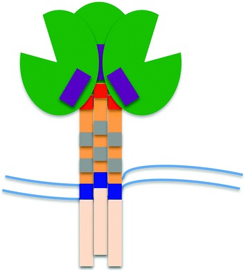
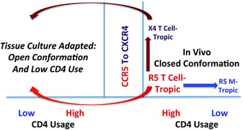
The surface envelope protein of any virus is major determinant of the host cell that is infected and as a result a major determinant of viral pathogenesis. Retroviruses have a single surface protein named Env. It is a trimer of heterodimers and is responsible for binding to the host cell receptor and mediating fusion between the viral and host membranes. In this review we will discuss the history of the discovery of the avian leukosis virus (ALV) and human immunodeficiency virus type 1 (HIV-1) Env proteins and their receptor specificity, comparing the many differences but having some similarities. Much of the progress in these fields has relied on viral genetics and genetic polymorphisms in the host population. A special feature of HIV-1 is that its persistent infection in its human host, to the point of depleting its favorite target cells, allows the virus to evolve new entry phenotypes to expand its host range into several new cell types. This variety of entry phenotypes has led to confusion in the field leading to the major form of entry phenotype of HIV-1 being overlooked until recently. Thus an important part of this story is the description and naming of the most abundant entry form of the virus: R5 T cell-tropic HIV-1.
Figures
Similar articles
-
Residues 28 to 39 of the Extracellular Loop 1 of Chicken Na+/H+ Exchanger Type I Mediate Cell Binding and Entry of Subgroup J Avian Leukosis Virus.J Virol. 2017 Dec 14;92(1):e01627-17. doi: 10.1128/JVI.01627-17. Print 2018 Jan 1. J Virol. 2017. PMID: 29070685 Free PMC article.
-
Low pH is required for avian sarcoma and leukosis virus Env-dependent viral penetration into the cytosol and not for viral uncoating.J Virol. 2004 Oct;78(19):10433-41. doi: 10.1128/JVI.78.19.10433-10441.2004. J Virol. 2004. PMID: 15367609 Free PMC article.
-
The viral envelope is a major determinant for the induction of lymphoid and myeloid tumours by avian leukosis virus subgroups A and J, respectively.J Gen Virol. 2002 Oct;83(Pt 10):2553-2561. doi: 10.1099/0022-1317-83-10-2553. J Gen Virol. 2002. PMID: 12237439
-
Alpharetrovirus envelope-receptor interactions.Curr Top Microbiol Immunol. 2003;281:107-36. doi: 10.1007/978-3-642-19012-4_3. Curr Top Microbiol Immunol. 2003. PMID: 12932076 Review.
-
Cellular entry of retroviruses.Adv Exp Med Biol. 2013;790:128-49. doi: 10.1007/978-1-4614-7651-1_7. Adv Exp Med Biol. 2013. PMID: 23884589 Review.
Cited by
-
Selective use of primate CD4 receptors by HIV-1.PLoS Biol. 2019 Jun 10;17(6):e3000304. doi: 10.1371/journal.pbio.3000304. eCollection 2019 Jun. PLoS Biol. 2019. PMID: 31181085 Free PMC article.
-
Isolation and characterization of a recombinant avian leukosis virus subgroup E from a commercial layer farm in Eastern China.Poult Sci. 2025 May;104(5):105053. doi: 10.1016/j.psj.2025.105053. Epub 2025 Mar 15. Poult Sci. 2025. PMID: 40132316 Free PMC article.
-
HIV replication and latency in monocytes and macrophages.Semin Immunol. 2021 Jan;51:101472. doi: 10.1016/j.smim.2021.101472. Epub 2021 Feb 27. Semin Immunol. 2021. PMID: 33648815 Free PMC article. Review.
-
Glycan masking in vaccine design: Targets, immunogens and applications.Front Immunol. 2023 Mar 23;14:1126034. doi: 10.3389/fimmu.2023.1126034. eCollection 2023. Front Immunol. 2023. PMID: 37033915 Free PMC article. Review.
-
Immunopathological investigation and genetic evolution of Avian leukosis virus Subgroup-J associated with myelocytomatosis in broiler flocks in Egypt.Virol J. 2024 Apr 10;21(1):83. doi: 10.1186/s12985-024-02329-7. Virol J. 2024. PMID: 38600532 Free PMC article.
References
-
- Arrildt KT, LaBranche CC, Joseph SB, Dukhovlinova EN, Graham WD, Ping L-H, Schnell G, Sturdevant CB, Kincer LP, Mallewa M, Heyderman RS, Van Rie A, Cohen MS, Spudich S, Price RW, Montefiori DC, Swanstrom R (2015) Phenotypic correlates of HIV-1 macrophage tropism. J Virol 89(22):11294–11311. doi:10.1128/jvi.00946-15 - PMC - PubMed
-
- Bates P, Young JA, Varmus HE (1993) A receptor for subgroup A Rous sarcoma virus is related to the low density lipoprotein receptor. Cell 74(6):1043–1051 - PubMed
-
- Bova CA, Manfredi JP, Swanstrom R (1986) Env genes of avian retroviruses: nucleotide sequence and molecular recombinants define host range determinants. Virology 152(2):343–354 - PubMed
Publication types
MeSH terms
Substances
LinkOut - more resources
Full Text Sources
Other Literature Sources
