. 2017 Jul 26:60:134-141.
doi: 10.1016/j.jbiomech.2017.06.031. Epub 2017 Jun 27.

Biomechanical properties of murine TMJ articular disc and condyle cartilage via AFM-nanoindentation

Affiliations

Biomechanical properties of murine TMJ articular disc and condyle cartilage via AFM-nanoindentation

Prashant Chandrasekaran et al. J Biomech. .

Abstract

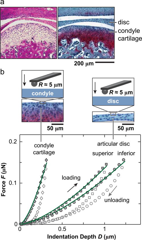
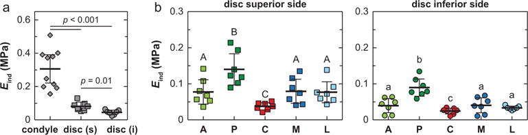
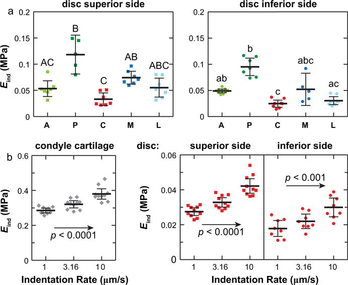
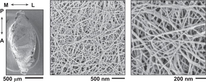
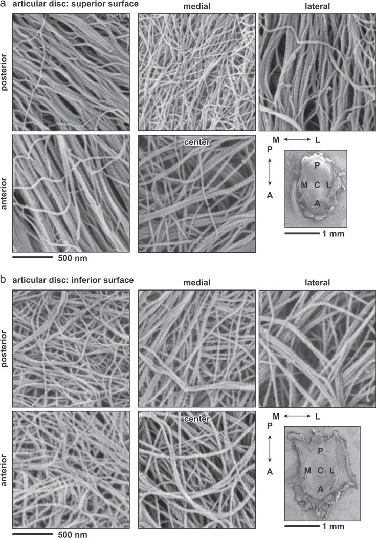
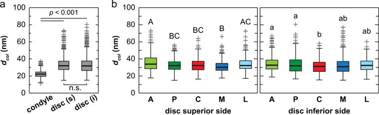
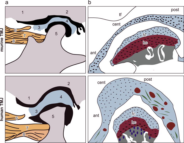
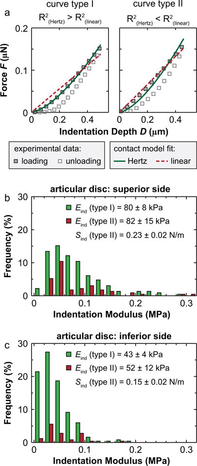
This study aims to quantify the biomechanical properties of murine temporomandibular joint (TMJ) articular disc and condyle cartilage using AFM-nanoindentation. For skeletally mature, 3-month old mice, the surface of condyle cartilage was found to be significantly stiffer (306±84kPa, mean±95% CI) than those of the superior (85±23kPa) and inferior (45±12kPa) sides of the articular disc. On the disc surface, significant heterogeneity was also detected across multiple anatomical sites, with the posterior end being the stiffest and central region being the softest. Using SEM, this study also found that the surfaces of disc are composed of anteroposteriorly oriented collagen fibers, which are sporadically covered by thinner random fibrils. Such fibrous nature results in both an F-D3/2 indentation response, which is a typical Hertzian response for soft continuum tissue under a spherical tip, and a linear F-D response, which is typical for fibrous tissues, further signifying the high degree of tissue heterogeneity. In comparison, the surface of condyle cartilage is dominated by thinner, randomly oriented collagen fibrils, leading to Hertzian-dominated indentation responses. As the first biomechanical study of murine TMJ, this work will provide a basis for future investigations of TMJ tissue development and osteoarthritis in various murine TMJ models.

Keywords: Fibrocartilage; Heterogeneity; Murine models; Nanoindentation; Temporomandibular joint.


Conflict of interest statement

Conflict of interest statement

The authors of this study have no personal or financial conflicts of interest with this work. All authors were fully involved in the study and preparation of this manuscript and the material within has not been and will not be submitted for publication elsewhere.

Figures

Fig. 1
Fig. 1
(a) Hematoxylin & Eosin (left) and Safranin-O/Fast Green (right) histology of sagittal sections showed the morphology of murine TMJ, highlighting the tissues of interest in this study: articular disc and mandibular condyle cartilage. (b) Representative indentation force versus depth, F-D, curves measured on the central regions of articular disc and mandibular condyle surfaces at 10 μm/s rate. The symbols are experimental data (density reduced for clarity), and solid lines are associated Hertz model fits to each of the loading F-D curve.
Fig. 2
Fig. 2
Effective indentation modulus, Eind, of 3-month old murine TMJ articular disc and condyle cartilage surfaces (n = 7 for each side of disc, and n = 10 for condyle cartilage, measured at 10 μm/s rate). (a) Comparison across three tissue sites. For condyle cartilage, each data point represents averaged values of ≥10 locations measured on the central region of one animal. For articular disc, each data point represents averaged value of ≥40 locations measured on all five regions of each animal. (b) Comparison across five different anatomical regions on the articular disc surface (A: anterior, P: posterior, C: central, M: medial, L: lateral), each data point represents averaged value of ≥8 locations measured on each region of one animal. Regions with different letters are statistically different (p < 0.05, mean ± 95% CI).
Fig. 3
Fig. 3
Heterogeneity and rate-dependence of Eind within individual animal. (a) Variation of Eind on the superior and inferior surfaces of TMJ articular disc from one mouse measured at 10 μm/s rate. (b) Rate-dependence of Eind measured on the central regions of TMJ condyle cartilage and articular disc from one mouse. Each data point represents one indentation location (≥8 locations measured on each region of one animal, p < 0.05, mean ± 95% CI).
Fig. 4
Fig. 4
SEM images of the central region of the mandibular condyle cartilage surface. The other four regions have similar fibrillar structure, and thus, are not shown.
Fig. 5
Fig. 5
SEM images of five different anatomical regions on (a) superior and (b) inferior sides of the articular disc surface.
Fig. 6
Fig. 6
Distribution of collagen fibril diameters measured on the surfaces of TMJ articular disc and mandibular condyle cartilage. (a) Comparison across the three tissue sites (for each disc surface, measurements were pooled from all five different regions), (b) Comparison across five different anatomical regions on the articular disc surface (A: anterior, P: posterior, C: central, M: medial, L: lateral) (≥300 fibrils from n = 3 animals for each region, regions with different letters are statistically different, p < 0.05, mean ± 95% CI).
Fig. 7
Fig. 7
Schematics of the anatomical structure of murine versus human TMJs. (a) Anatomical structure of the TMJ: 1. articular eminence of temporal bone, 2. glenoid fossa of temporal bone, 3. anterior band of the articular disc, 4. posterior band of the articular disc, 5. mandibular condyle, 6. upper head of lateral pterygoid muscle, 7. upper head of the lateral pterygoid muscle connected to the mandible and 8. lower head of the lateral pterygoid muscle connected to the mandible condyle (adapted from (Suzuki and Iwata (2016)) with permission). (b) More detailed schematics of TMJ disc and condyle cartilage structure. In the murine TMJ, the articular disc has substantially smaller thickness compared to the condyle cartilage. This contrast is absent in larger animal TMJs (abbreviations, tf: temporal fossa, ant: anterior end, cent: central region, post: posterior end, fc: fibrocartilage, hc: hyaline cartilage). The tissue thickness values are: human: TMJ disc ~0.5–3 mm, condyle fibrocartilage layer: 400–600 μm, hyaline cartilage layer ~300–600 μm, as estimated in Bibb et al. (1993), Wright et al. (2016); murine TMJ disc ~30–60 μm, condyle fibrocartilage layer ~25–40 μm, hyaline cartilage layer ~90–120 μm.
Fig. 8
Fig. 8
Non-Hertzian indentation responses of the TMJ articular disc surface. (a) Two types of representative indentation force versus depth F-D curves from the posterior region of the TMJ disc superior surface. Type I: Hertzian indentation curve where FD3/2, where R2 (Hertz) > R2 (linear). Type II: linear indentation curve where FD, where R2 (Hertz) < R2 (linear). (b, c) Histograms of the distribution of Eind from two indentation curve types on (b) the superior and (c) the inferior surfaces of the TMJ disc (≥300 locations from n = 7 animals for each side of the disc measured at 10 μm/s rate. No significant difference in Eind was found between type I and II curves on each surface).

Similar articles

Cited by

References

    1. Allen KD, Athanasiou KA. Viscoelastic characterization of the porcine temporomandibular joint disc under unconfined compression. J Biomech. 2006;39:312–322. - PubMed
    1. Batista MA, Nia HT, Önnerfjord P, Cox KA, Ortiz C, Grodzinsky AJ, Heinegård D, Han L. Nanomechanical phenotype of chondroadherin-null murine articular cartilage. Matrix Biol. 2014;38:84–90. - PMC - PubMed
    1. Beatty MW, Bruno MJ, Iwasaki LR, Nickel JC. Strain rate dependent orthotropic properties of pristine and impulsively loaded porcine temporomandibular joint disk. J Biomed Mater Res. 2001;57:25–34. - PubMed
    1. Bechtold TE, Saunders C, Mundy C, Um H, Decker RS, Salhab I, Kurio N, Billings PC, Pacifici M, Nah HD, Koyama E. Excess BMP signaling in heterotopic cartilage forming in Prg4-null TMJ discs. J Dent Res. 2016;95:292–301. - PMC - PubMed
    1. Bertram S, Rudisch A, Innerhofer K, Pumpel E, Grubwieser G, Emshoff R. Diagnosing TMJ internal derangement and osteoarthritis with magnetic resonance imaging. J Am Dent Assoc. 2001;132:753–761. - PubMed

MeSH terms

Substances