

Inter- and Intra-Observer Variability of the Volume of Cervical Ossification of the Posterior Longitudinal Ligament Using Medical Image Processing Software
- PMID: 28689393
- PMCID: PMC5544367
- DOI: 10.3340/jkns.2015.0708.014
Inter- and Intra-Observer Variability of the Volume of Cervical Ossification of the Posterior Longitudinal Ligament Using Medical Image Processing Software
Abstract
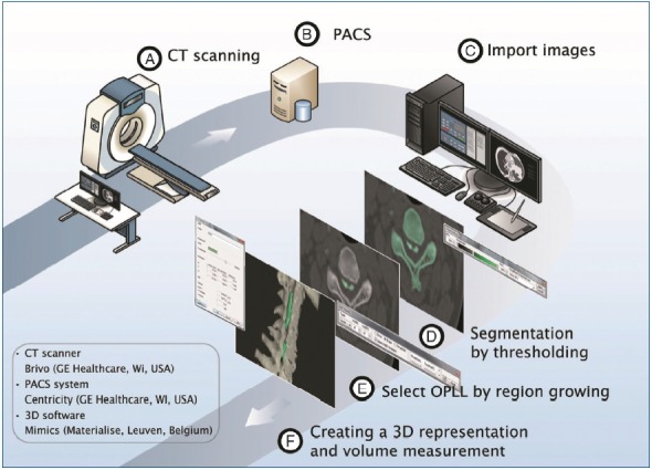
Objective: Computed tomography (CT)-based method of three dimensional (3D) analysis (MIMICS®, Materialise, Leuven, Belgium) is reported as very useful software for evaluation of OPLL, but its reliability and reproducibility are obscure. This study was conducted to evaluate the accuracy of MIMICS® system, and inter- and intra-observer reliability in the measurement of OPLL.
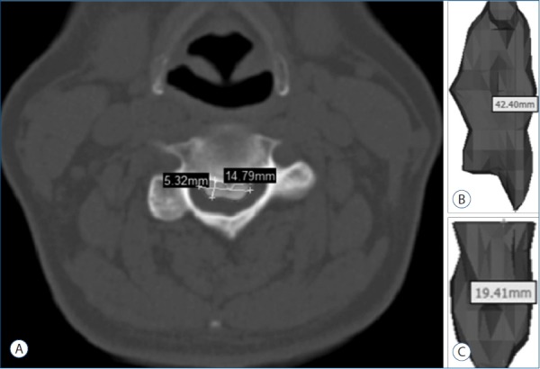
Methods: Three neurosurgeons independently analyzed the randomly selected 10 OPLL cases with medical image processing software (MIMICS®) which create 3D model with Digital Imaging and Communication in Medicine (DICOM) data from CT images after brief explanation was given to examiners before the image construction steps. To assess the reliability of inter- and intra-examiner intraclass correlation coefficient (ICC), 3 examiners measured 4 parameters (volume, length, width, and length) in 10 cases 2 times with 1-week interval.
Results: The inter-examiner ICCs among 3 examiners were 0.996 (95% confidence interval [CI], 0.987-0.999) for volume measurement, 0.973 (95% CI, 0.907-0.978) for thickness, 0.969 (95% CI, 0.895-0.993) for width, and 0.995 (95% CI, 0.983-0.999) for length. The intra-examiner ICCs were 0.994 (range, 0.991-0.996) for volume, 0.996 (range, 0.944-0.998) for length, 0.930 (range, 0.873-0.947) for width, and 0.987 (range, 0.985-0.995) for length.
Conclusion: The medical image processing software (MIMICS®) provided detailed quantification OPLL volume with minimal error of inter- and intra-observer reliability in the measurement of OPLL.
Keywords: 3D analysis; MIMICS; Ossification; Posterior longitudinal ligament; Volume.
Figures
Similar articles
-
Inter- and intra-observer variability of a cervical OPLL classification using reconstructed CT images.Clin Orthop Surg. 2010 Mar;2(1):8-12. doi: 10.4055/cios.2010.2.1.8. Epub 2010 Feb 4. Clin Orthop Surg. 2010. PMID: 20190995 Free PMC article.
-
Usefulness of 3-dimensional Measurement of Ossification of the Posterior Longitudinal Ligament (OPLL) in Patients With OPLL-induced Myelopathy.Spine (Phila Pa 1976). 2015 Oct 1;40(19):1479-86. doi: 10.1097/BRS.0000000000001072. Spine (Phila Pa 1976). 2015. PMID: 26208225
-
Effect of posterior instrumented fusion on three-dimensional volumetric growth of cervical ossification of the posterior longitudinal ligament: a multiple regression analysis.Spine J. 2018 Oct;18(10):1779-1786. doi: 10.1016/j.spinee.2018.03.002. Epub 2018 Mar 8. Spine J. 2018. PMID: 29526640
-
Three-dimensional evaluation of volume change in ossification of the posterior longitudinal ligament of the cervical spine using computed tomography.Eur Spine J. 2013 Nov;22(11):2569-74. doi: 10.1007/s00586-013-2989-9. Epub 2013 Sep 3. Eur Spine J. 2013. PMID: 24000076 Free PMC article.
-
Computer-assisted measurement of the size of ossification in patients with ossification of the posterior longitudinal ligament in the cervical spine.J Orthop Sci. 2005 Sep;10(5):451-6. doi: 10.1007/s00776-005-0925-5. J Orthop Sci. 2005. PMID: 16193355
Cited by
-
Three-dimensional imaging analysis for the diagnosis of dural ossification in thoracic ossification of the ligamentum flavum: a multicenter study.Quant Imaging Med Surg. 2023 Jan 1;13(1):417-427. doi: 10.21037/qims-22-418. Epub 2022 Nov 14. Quant Imaging Med Surg. 2023. PMID: 36620130 Free PMC article.
-
The clinical value of three-dimensional measurement in the diagnosis of thoracic myelopathy caused by ossification of the ligamentum flavum.Quant Imaging Med Surg. 2021 May;11(5):2040-2051. doi: 10.21037/qims-20-713. Quant Imaging Med Surg. 2021. PMID: 33936985 Free PMC article.
-
Evaluation of Vertebral Function and Long-Term Quality of Life after Percutaneous Minimally Invasive Surgery in Patients with Thoracolumbar Spine Fractures.Comput Math Methods Med. 2022 Feb 23;2022:2723542. doi: 10.1155/2022/2723542. eCollection 2022. Comput Math Methods Med. 2022. Retraction in: Comput Math Methods Med. 2023 Sep 27;2023:9859432. doi: 10.1155/2023/9859432. PMID: 35251297 Free PMC article. Retracted. Clinical Trial.
-
A feasibility study of modified self-efficacy for the improvement of adverse emotions and quality of life in traumatic fracture patients.Am J Transl Res. 2021 Jun 15;13(6):6507-6515. eCollection 2021. Am J Transl Res. 2021. PMID: 34306391 Free PMC article.
-
Accuracy and Reliability of Computer-aided Anatomical Measurements for Vertebral Body and Disc Based on Computed Tomography Scans.Orthop Surg. 2020 Aug;12(4):1182-1189. doi: 10.1111/os.12729. Epub 2020 Jul 3. Orthop Surg. 2020. PMID: 32618427 Free PMC article.
References
-
- Chiba K, Kato Y, Tsuzuki N, Nagata K, Toyama Y, Iwasaki M, et al. Computer-assisted measurement of the size of ossification in patients with ossification of the posterior longitudinal ligament in the cervical spine. J Orthop Sci. 2005;10:451–456. - PubMed
-
- Chiba K, Ogawa Y, Ishii K, Takaishi H, Nakamura M, Maruiwa H, et al. Long-term results of expansive open-door laminoplasty for cervical myelopathy--average 14-year follow-up study. Spine (Phila Pa 1976) 2006;31:2998–3005. - PubMed
-
- Chiba K, Yamamoto I, Hirabayashi H, Iwasaki M, Goto H, Yonenobu K, et al. Multicenter study investigating the postoperative progression of ossification of the posterior longitudinal ligament in the cervical spine: a new computer-assisted measurement. J Neurosurg Spine. 2005;3:17–23. - PubMed
LinkOut - more resources
Full Text Sources
Other Literature Sources
