Review
. 2018 Sep;118(Pt B):54-67.
doi: 10.1016/j.neuropsychologia.2017.07.003. Epub 2017 Jul 8.

The anatomy of apathy: A neurocognitive framework for amotivated behaviour

Affiliations
Review

The anatomy of apathy: A neurocognitive framework for amotivated behaviour

C Le Heron et al. Neuropsychologia. 2018 Sep.

Abstract

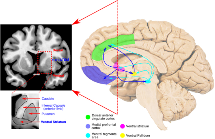
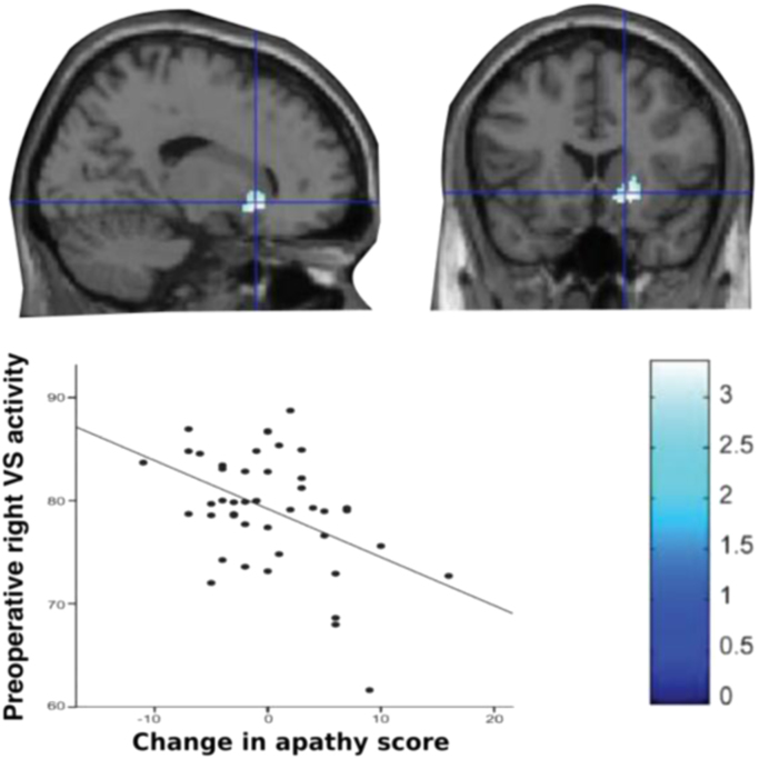
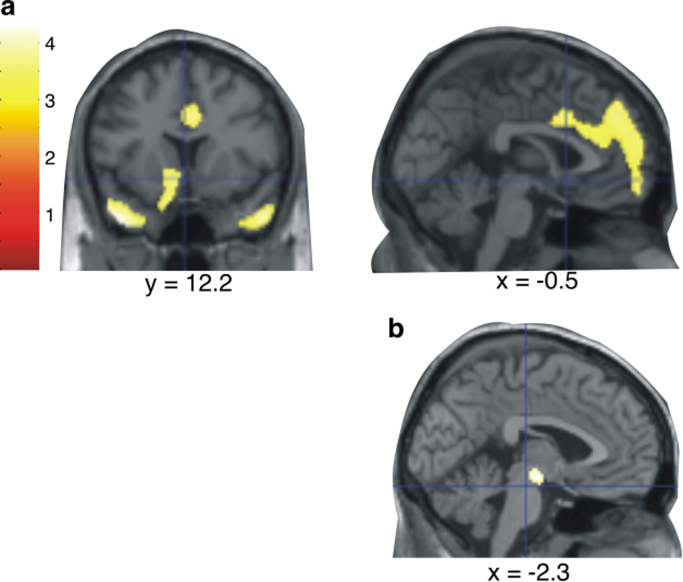
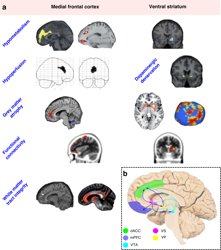
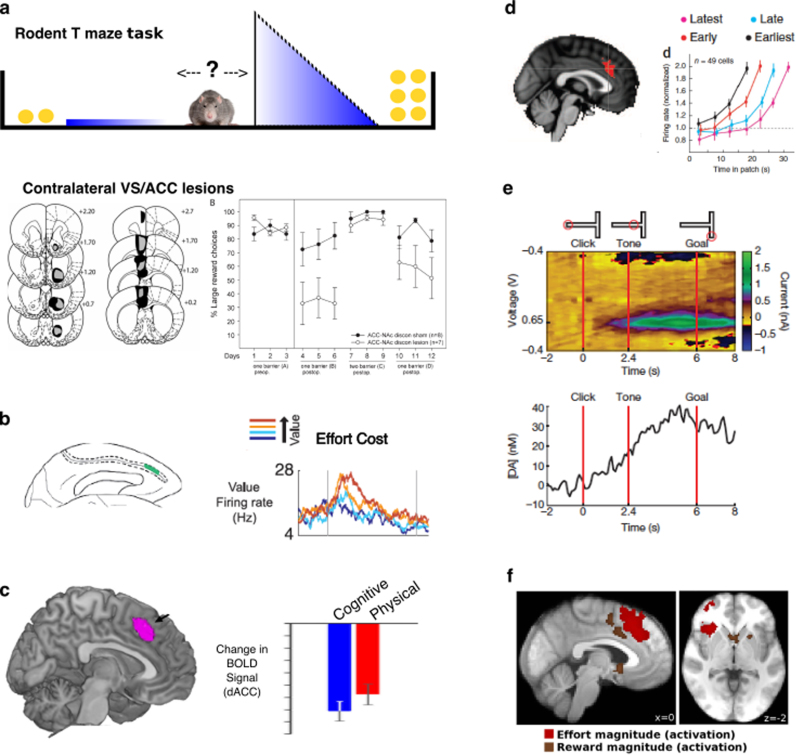
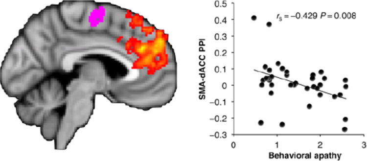
Apathy is a debilitating syndrome associated with many neurological disorders, including several common neurodegenerative diseases such as Parkinson's disease and Alzheimer's disease, and focal lesion syndromes such as stroke. Here, we review neuroimaging studies to identify anatomical correlates of apathy, across brain disorders. Our analysis reveals that apathy is strongly associated with disruption particularly of dorsal anterior cingulate cortex (dACC), ventral striatum (VS) and connected brain regions. Remarkably, these changes are consistent across clinical disorders and imaging modalities. Review of the neuroimaging findings allows us to develop a neurocognitive framework to consider potential mechanisms underlying apathy. According to this perspective, an interconnected group of brain regions - with dACC and VS at its core - plays a crucial role in normal motivated behaviour. Specifically we argue that motivated behaviour requires a willingness to work, to keep working, and to learn what is worth working for. We propose that deficits in any one or more of these processes can lead to the clinical syndrome of apathy, and outline specific approaches to test this hypothesis. A richer neurobiological understanding of the mechanisms underlying apathy should ultimately facilitate development of effective therapies for this disabling condition.

Keywords: Anterior cingulate cortex; Apathy; Decision making; Motivation; Reward; Ventral striatum.


Figures

Fig. 1
Fig. 1
A reciprocally connected network of brain regions for normal motivated behaviour. A network of medial frontal and striatal regions has been strongly implicated in the generation of motivated behaviour in healthy people. Disruption of components of – or connections between – this network is strongly associated with apathy across brain disorders. The dorsal anterior cingulate cortex (dACC) encompasses the anterior cingulate sulcus (Brodmann area 24c & 32). Here medial prefrontal cortex refers to ventromedial prefrontal and orbitofrontal cortex, two anatomically overlapping regions (particularly Brodmann areas 10, 11 and 47) – see Neubert et al. (2015) for a detailed anatomical description.
Fig. 2
Fig. 2
Decreased metabolism within right ventral striatum is associated with greater increase in apathy scores following deep brain stimulation surgery.
Fig. 3
Fig. 3
Brain regions in which reduced metabolism was associated with apathy, in a mixed group of patients with dementia (a). A disjunction analysis (controlling for other behavioural disturbances associated with altered metabolism) revealed apathy was specifically associated with hypometabolism in the region of the VTA (b).
Fig. 4
Fig. 4
Consistent anatomical associations with apathy across imaging modalities and underlying disorders. Changes affecting dorsal anterior cingulate cortex and ventral striatum are particularly prominent (a). A connected system of brain areas consistently associated with apathy, which form an interconnected network within the medial forebrain (b). dACC – dorsal anterior cingulate cortex; mPFC – medial prefrontal cortex; VTA – ventral tegmental area; VS – ventral striatum; VP – ventral pallidum. Images (from top left) adapted with permissions from: (Huang et al., 2013, Robert et al., 2014, Schroeter et al., 2011; Migneco et al., 2001, Remy et al., 2005, Stanton et al., 2013; Baggio et al., 2015, Bruen et al., 2008, Carriere et al., 2014; Hollocks et al., 2015, Marshall et al., 2007, Ota et al., 2012).
Fig. 5
Fig. 5
Importance of ACC and VS for motivated behaviour. Effort based decision making. Lesions to ACC, VS or crossed ACC/VS disconnection lesions (pictured) cause rodents to choose more low effort/low reward options, without altering their preference for high rewards options if effort costs are equal (a) (from Hauber and Sommer, 2009). Single neurons within macaque ACC modulate firing rates to the net value of a choice (reward – effort) (b) (from Kennerley et al., 2009). BOLD activity within human ACC signals the subjective value of a choice (reward – effort), whether the cost is physical or cognitive (c) (from Chong et al., 2017) Modulating behavior to foreground and background environment characteristics. BOLD activity correlates with the value of foraging (switching from current option), rather than the value of the actual chosen option (d – left panel) (from Kolling et al., 2012). Single neuronal recordings from the ACC of macaques performing a patch leaving foraging task show earlier increases in firing rate on trials where the animal leaves the patch faster (d – right panel) (from Hayden et al., 2011b) Sustaining behavior towards a goal. Recordings from rodent VS show ramping dopamine signals as the animal approaches a rewarding outcome, thought to drive persistence of behaviour towards the goal (e) (from Howe et al., 2013) Monitoring outcomes of actions. ACC and VS activity encodes information on both reward and effort magnitudes during the outcome phase of an action (f) (from Scholl et al., 2015) (all figures reproduced with permissions).
Fig. 6
Fig. 6
Level of functional connectivity between dorsal ACC (yellow-orange) and supplementary motor area (purple) predicts behavioural apathy scores in healthy subjects (higher score corresponds to greater apathy traits) (fromBonnelle et al., 2016with permission).

References

    1. Aarsland D., Marsh L., Schrag A. Neuropsychiatric symptoms in Parkinson's disease. Mov. Disord. 2009 - PMC - PubMed
    1. Adam R., Leff A., Sinha N., Turner C., Bays P., Draganski B., Husain M. Dopamine reverses reward insensitivity in apathy following globus pallidus lesions. Cortex. 2013;49:1292–1303. - PMC - PubMed
    1. Agosta F., Galantucci S., Svetel M., Lukić M.J., Copetti M., Davidovic K., Tomić A., Spinelli E.G., Kostić V.S., Filippi M. Clinical, cognitive, and behavioural correlates of white matter damage in progressive supranuclear palsy. J. Neurol. 2014;261:913–924. - PubMed
    1. Alexander G.E., Crutcher M.D. Functional architecture of basal ganglia circuits - neural substrates of parallel processing. Trends Neurosci. 1990;13:266–271. - PubMed
    1. Ang Y.-S., Lockwood P., Apps M.A.J., Muhammed K., Husain M. Distinct Subtypes of Apathy Revealed by the Apathy Motivation Index. PLoS One. 2017;12:e0169938. - PMC - PubMed

Publication types