. 2017 Aug 8;9(2):587-599.
doi: 10.1016/j.stemcr.2017.06.005. Epub 2017 Jul 6.

MAPT Genetic Variation and Neuronal Maturity Alter Isoform Expression Affecting Axonal Transport in iPSC-Derived Dopamine Neurons

Affiliations

MAPT Genetic Variation and Neuronal Maturity Alter Isoform Expression Affecting Axonal Transport in iPSC-Derived Dopamine Neurons

Joel E Beevers et al. Stem Cell Reports. .

Abstract

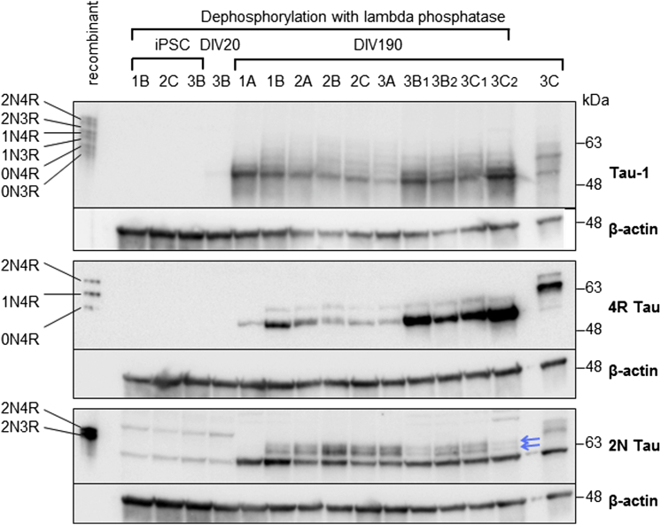
The H1 haplotype of the microtubule-associated protein tau (MAPT) locus is genetically associated with neurodegenerative diseases, including Parkinson's disease (PD), and affects gene expression and splicing. However, the functional impact on neurons of such expression differences has yet to be fully elucidated. Here, we employ extended maturation phases during differentiation of induced pluripotent stem cells (iPSCs) into mature dopaminergic neuronal cultures to obtain cultures expressing all six adult tau protein isoforms. After 6 months of maturation, levels of exon 3+ and exon 10+ transcripts approach those of adult brain. Mature dopaminergic neuronal cultures display haplotype differences in expression, with H1 expressing 22% higher levels of MAPT transcripts than H2 and H2 expressing 2-fold greater exon 3+ transcripts than H1. Furthermore, knocking down adult tau protein variants alters axonal transport velocities in mature iPSC-derived dopaminergic neuronal cultures. This work links haplotype-specific MAPT expression with a biologically functional outcome relevant for PD.

Keywords: MAPT; Parkinson's disease; dopamine neurons; iPSC; tau.


Figures

Figure 1
Figure 1
Differentiation of Induced Pluripotent Stem Cells with MAPT H1/H2 Genotype into Dopaminergic Neuronal Cultures (A) Genotyping PCR distinguishes the 238 bp indel in MAPT intron 9, showing the presence of both the H1 and H2 alleles in all eight iPSC clones. Clones are identified by the number of the individual (1–3) then by the clone generated from reprogramming of the fibroblasts of that individual (A–C). (B) Western blots showing iPSC differentiation into dopamine neuronal cultures. Neuronal marker β3-tubulin (TUJ1) (expressed by DIV20) and tyrosine hydroxylase (TH) are shown at DIV190. Samples from two differentiations are identified by a “1” or “2” suffix. (C) Efficiency of dopaminergic differentiation was quantified from at least two clones per individual. Individuals differentiated with similar efficiencies into neurons determined by TUJ1/DAPI. The proportion of neurons that were dopaminergic was determined by TH/TUJ1. Mean ± SEM of n = 2 or 3 clones per individual; 4 images per clone. (D) Immunofluorescent co-labelling of differentiated neuronal cultures from clone 3B fixed at DIV27 (1 week after re-plating) demonstrates expression of dopaminergic neuronal protein TH together with β3-tubulin. Scale bars, 50 μm. See also Figures S1 and S2.
Figure 2
Figure 2
MAPT Adult Isoforms Increase in Expression over a 6-Month Differentiation (A–C) Real-time qPCR analysis demonstrated changes in MAPT expression over 24 weeks of maturation to DIV188. Each graph shows mean ± SEM, n = 7 clones (all clones except 1A). At DIV188, peak mean inclusion for the alternatively spliced exons 3 and 10 was 5.6% (B) and 18.2% (C), respectively. For each graph (A–C), linear regression performed using all data points from DIV48 to DIV188 with an F test confirming that each slope is significantly different from zero: (A) y = −0.01303 × x + 0.7279, F = 4.615, p = 0.0391; (B) y = 0.2553 × x − 0.7078, F = 23.32, p < 0.0001; (C) y = 0.7416 × x − 0.05151, F = 96.28, p < 0.0001. (D) Exon inclusion from TaqMan-based real-time qPCR expression assays performed on human midbrain cDNA. Mean ± SEM, n = 5 individuals; exon 3+ mean = 6.79 ± 1.43; exon 10+ mean = 35.22 ± 2.43. See also Figure S3.
Figure 3
Figure 3
All Major Tau Protein Isoforms Are Expressed in iPSC-Derived Dopaminergic Neuronal Cultures Differentiated for 6 Months Western blots showing the presence of all six major isoforms of mature tau protein in dopaminergic neuronal cultures at DIV190, as detected by antibodies against total tau (Tau-1) or specific isoforms (4R Tau, 2N Tau). All samples dephosphorylated, except the right lane as an untreated control. 2N Tau blot: central bands indicated by blue arrows represent the two 2N Tau isoforms. Additional bands are considered non-specific as they appear in the iPSC lysate in which no tau is detectable.
Figure 4
Figure 4
Dopaminergic Neuronal Cultures Exhibit Significant Differences in Isoform Expression from H1 and H2 Alleles at 6 Months TaqMan-based real-time qPCR expression assays on samples at DIV190. Mean ± SEM; individual 1, n = 1 clone; individuals 2 and 3, n = 3 clones; ≥3 cDNA samples per clone. (A) Total MAPT expression reported as relative ΔCT of geometric mean of three housekeeper genes (GAPDH, HPRT1, and ACTB). n.s., not significant, one-way ANOVA. (B) Percent inclusion of alternatively spliced exons 3 (light blue) and 10 (dark blue) at DIV190. The three individuals show similar inclusion of exon 3, whereas individual 3 shows a significantly greater inclusion of exon 10. Significant difference between groups in an unpaired t test: ∗∗∗p = 0.0005. (C–E) Allele-specific expression assays distinguishing transcripts of H1 and H2 allelic origin, presented as H1:H2 ratio, i.e., values >1 show higher H1 expression. Data from analysis of human midbrain (C) n = 9; (D) n = 5; and (E) n = 9. (C) Individuals 2 and 3 show significantly greater expression of total MAPT transcripts from the H1 chromosome (individual 2, ∗∗p = 0.0059; individual 3, p = 0.0471; midbrain, n.s.). (D and E) The H1:H2 ratio is normalized to the H1:H2 ratio of total MAPT transcripts per sample. Significant difference from mean of 1 in a one-sample t test. (D) There are 2-fold greater exon 3-containing transcripts coming from the H2 chromosome. Individual 2, ∗∗p = 0.0032; individual 3, ∗∗p = 0.0040; midbrain, ∗∗p = 0.0020. (E) Haplotype-specific expression of exon 10 varies between individuals and midbrain: Individual 2, n.s.; individual 3, ∗∗∗p = 0.0008; midbrain, ∗∗p = 0.0005. Significant difference between groups in an unpaired t test: ∗∗∗p = 0.0005. See also Figures S3 and S4.
Figure 5
Figure 5
A Deletion in MAPT Intron 10 Decreases Binding of Factors that Regulate Exon 10 Splicing (A) Sequencing chromatograms for MAPT intron 10 showing the expected sequence for genomic DNA (both H1 and H2 alleles together) for individuals 1 and 2. The red x's represent divergent chromatograms from two overlapping sequences caused by an indel on one allele for individual 3. (B) Sequencing chromatograms for MAPT intron 10 showing single allelic genomic DNA from individual 3. The H2 allele showed a ΔCTT variation. (C) Electrophoretic mobility shift assay using an RNA probe for an intron 10 wild-type (WT) sequence (lanes 1–6) and an RNA probe for an intron 10 ΔCTT variant sequence (lanes 7–12), with and without competitors. (D) Western blot of RNA-protein pull-down using the WT and ΔCTT RNA probes. Lanes indicate proteins obtained from nuclear extract only (NE), beads only (B), NE and B (NEB), WT RNA probe and the ΔCTT probe. Blots are shown for PTPB1 and RMB4. See also Figure S5.
Figure 6
Figure 6
Knockdown of 4R Tau Increases Velocity of Mitochondrial Axonal Transport in iPSC-Derived Dopaminergic Neuronal Cultures Differentiated for 5 Months (A) Western blots showing knockdown of tau protein in dopaminergic neuronal cultures 4 weeks post-transduction. p = 0.0457. (B) Western blots after protein dephosphorylation show knockdown of tau protein and 4R tau isoforms persisting in 5-month dopaminergic neuronal cultures with β-actin loading control. (C) Fluorescence microscopy image of EBFP2 expression during axonal transport live imaging at 5 months. Scale bar, 25 μm. (D) Selected time-lapse fluorescence microscopy images of axonal mitochondria in dopaminergic neuronal cultures stained with MitoTracker Deep Red. Arrowheads show motile mitochondria. (E) Kymograph time-space plot of a trace along the linear path of an axon, from which mitochondrial motility parameters can be determined. (F) Cumulative frequency graphs of average mitochondrial velocity in dopaminergic neuronal cultures at (top) 4 weeks post-transduction and (bottom) 5 months post-transduction. (Left) Average total velocity of each measured motile mitochondrion; (right) average velocity of each measured motile mitochondrion after removal of time when paused. All four graphs were statistically significant in Kruskall-Wallis nonparametric tests: upper left, p = 0.0033; upper right, p = 0.0200; lower left, p < 0.0001; lower right, p < 0.0001. Significance in the follow-up Dunn's multiple comparisons test (nonparametric) compared with scrambled shRNA control: p < 0.05; ∗∗∗p < 0.001. See also Figure S6.

References

    1. Allen M., Kachadoorian M., Quicksall Z., Zou F., Chai H.S., Younkin C., Crook J.E., Pankratz V.S., Carrasquillo M.M., Krishnan S. Association of MAPT haplotypes with Alzheimer's disease risk and MAPT brain gene expression levels. Alzheimers Res. Ther. 2014;6:39. - PMC - PubMed
    1. Andreadis A., Brown W.M., Kosik K.S. Structure and novel exons of the human tau gene. Biochemistry. 1992;31:10626–10633. - PubMed
    1. Arai T., Ikeda K., Akiyama H., Shikamoto Y., Tsuchiya K., Yagishita S., Beach T., Rogers J., Schwab C., McGeer P.L. Distinct isoforms of tau aggregated in neurons and glial cells in brains of patients with Pick's disease, corticobasal degeneration and progressive supranuclear palsy. Acta Neuropathol. 2001;101:167–173. - PubMed
    1. Buee Scherrer V., Hof P.R., Buee L., Leveugle B., Vermersch P., Perl D.P., Olanow C.W., Delacourte A. Hyperphosphorylated tau proteins differentiate corticobasal degeneration and Pick's disease. Acta Neuropathol. 1996;91:351–359. - PubMed
    1. Caffrey T.M., Joachim C., Paracchini S., Esiri M.M., Wade-Martins R. Haplotype-specific expression of exon 10 at the human MAPT locus. Hum. Mol. Genet. 2006;15:3529–3537. - PubMed

Publication types