. 2017:2017:7150376.
doi: 10.1155/2017/7150376. Epub 2017 Jun 13.

Novel Therapeutic Effects of Leonurine On Ischemic Stroke: New Mechanisms of BBB Integrity

Affiliations

Novel Therapeutic Effects of Leonurine On Ischemic Stroke: New Mechanisms of BBB Integrity

Qiu-Yan Zhang et al. Oxid Med Cell Longev. 2017.

Abstract

Stroke is a leading cause of morbidity and mortality globally. Leonurine (also named SCM-198), a compound extracted from Herba leonuri, was effective on the prevention of various cardiovascular and brain diseases. The purpose of this study was to explore the possible therapeutic potential of SCM-198 against ischemia reperfusion injury and underlying mechanisms. In the in vivo transient middle cerebral artery occlusion (tMCAO) rat model, we found that treatment with SCM-198 could decrease infarct volume and improve neurological deficit by protecting against blood-brain barrier (BBB) breakdown. In the in vitro model of cell oxygen-glucose deprivation and reoxygenation (OGD/R), consistent results were obtained with decreased reactive oxygen species (ROS) production and maintained the BBB integrity. Further study demonstrated that SCM-198 increased the expression of histone deacetylase- (HDAC-) 4 which could inhibit NADPH oxidase- (NOX-) 4 and matrix metalloproteinase- (MMP-) 9 expression, resulting in the elevation of tight junction proteins, including claudin-5, occludin, and zonula occluden- (ZO-) 1. These results indicated SCM-198 protected BBB integrity by regulating the HDAC4/NOX4/MMP-9 tight junction pathway. Our findings provided novel insights into the protective effects and mechanisms of SCM-198 on ischemic stroke, indicating SCM-198 as a new class of potential drug against acute onset of ischemic stroke.


Figures
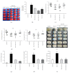
Figure 1
Figure 1
Posttreatment with SCM-198 significantly protected against the damage induced by tMCAO. The rats were subjected to 90 min MCAO before reperfusion. SCM-198 decreased the infarct volume and improved neurological scores. (a) Representative pictures of coronal sections from ischemic rat brain stained with TTC. (b) The quantitative analysis of the number of infarct size. F (4, 31) = 35.04, P < 0.0001. (c–e) Neurological function was ameliorated by SCM-198 after ischemia reperfusion. Values are expressed as mean ± SEM. #p < 0.05 versus the control group, p < 0.05 versus the tMCAO group, n = 8. Posttreatment with SCM-198 maintained BBB integrity. (f, g) At the end of treatment, the Evans blue dye was injected into the vein following 24 h circulation. (f) Representative pictures of coronal sections from rat brain stained with Evans blue dye after ischemic reperfusion. (g) The quantitative analysis of Evans blue leakage after ischemia reperfusion, F (3, 39) = 59.33, P < 0.0001. Edema volume ((h), F (3, 20) = 29.14, P < 0.0001) and water content ((i), F (3, 52) = 18.06, P < 0.0001) were decreased by SCM-198 after ischemia reperfusion. Values are expressed as mean ± SEM. #p < 0.05 versus the control group, p < 0.05 versus the tMCAO group, n = 6.
Figure 2
Figure 2
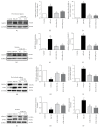
SCM-198 protected against TJ degradation induced by cerebral ischemic reperfusion. (a, d) Western blots of peri-ischemic region and striatum tissues in the tMCAO model showed increased the loss of occludin and ZO-1 levels, but reversed by SCM-198 treatment. GAPDH was used as the loading control. (b-c, e-f) The quantitative analysis occludin and ZO-1 levels were calculated and expressed relative to control. F (3, 12) = 18.65, P < 0.0001, Figure 2(b); F (3, 22) = 38.22, P < 0.0001, Figure 2(c); F (3, 8) = 8.228, P = 0.0079, Figure 2(e); F (3, 16) = 17.23, P < 0.0001, Figure 2(f). (g–j) Posttreatment with SCM-198 at 2 h and 0.5 h after reperfusion notably improved occludin and ZO-1 mRNA expression. SCM-198 revealed significantly protection. F (3, 6) = 25.11, P = 0.0009, Figure 2(g); F (3, 8) = 7.631, P = 0.0099, Figure 2(h); F (3, 8) = 10.04, P = 0.0044, Figure 2(i); F (3, 8) = 9.389, P = 0.0053, Figure 2(j). Values are expressed as mean ± SEM. #p < 0.05 versus the control group, p < 0.05 versus the tMCAO group, n = 4.
Figure 3
Figure 3
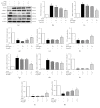
SCM-198 mediated the expression of MMP-9, NOX4, and HDAC4 in tMCAO rat. Reperfusion increased the loss of HDAC4 and the expression of MMP-9 and NOX4 in both peri-ischemic region and striatum tissues of the brain. (a–c) Reperfusion exacerbated the protein and mRNA expression of MMP-9 in peri-ischemic regions. SCM-198 significantly inhibited MMP-9 mRNA expression in the cortex. F (3, 9) = 38.73, P < 0.0001, Figure 3(b); F (3, 8) = 85.85, P < 0.0001, Figure 3(c). (d–f) Reperfusion improved the protein and mRNA expression of MMP-9 in the striatum. SCM-198 inhibited MMP-9 mRNA expression in the striatum. F (3, 10) = 14.65, P = 0.0005, Figure 3(e); F (3, 8) = 42.11, P < 0.0001, Figure 3(f); (g–l) Reperfusion increased the loss of HDAC4 and the expression of NOX4 in both peri-ischemic region and striatum tissues of the brain. GAPDH was used as the loading control. F (3, 15) = 51.79, P < 0.0001, Figure 3(h); F (3, 8) = 31, P < 0.0001, Figure 3(i); F (3, 11) = 24.47, P < 0.0001, Figure 3(k); F (3, 8) = 14.09, P = 0.0015, Figure 3(l). Values are expressed as mean ± SEM. #p < 0.05 versus the control group, p < 0.05 versus the tMCAO group, n = 4.
Figure 4
Figure 4
SCM-198 protected against ischemia-like injury in the in vitro BBB model. bEnd.3 cell line was exposed to 6 h of OGD followed by 4 h reperfusion after treated with different concentrations of SCM-198. (a) OGD/R markedly increased cell death, SCM-198 dose-dependently improved the cell viability, F (3, 14) = 12.57, P = 0.0003. (b) SCM-198 reduced the LDH leakage in the supernatant after OGD/R, F (3, 4) = 39.25, P = 0.002. (c) The fluorescence intensity of intracellular ROS, F (4, 12) = 43.35, P < 0.0001. (d) OGD/R produced abundant ROS in the cells, while SCM-198 significantly reduced the ROS formation, scale bar = 10 μm. Values are expressed as mean ± SEM. #p < 0.05 versus the control group, p < 0.05 versus the OGD group, n = 3.
Figure 5
Figure 5
SCM-198 reduced the degradation of TJs induced by OGD/R. (a) FITC-dextran was used to determine BBB integrity. After reperfusion the leakage of dextran in the plate was remarkably increased. SCM-198 could reduce the leakage, F (4, 10) = 6.671, P = 0.007. (b) Expressions of TJs, occludin, claudin-5, and ZO-1 were evaluated by western blot 4 h after reperfusion. OGD/R induced the obvious loss of TJs, and SCM-198 reversed this loss. (c–e) The results were quantified and expressed relative to control, F (4, 10) = 133.1, P < 0.0001, Figure 5(c); F (4, 14) = 26.32, P < 0.0001, Figure 5(d); F (4, 14) = 22.99, P < 0.0001, Figure 5(e). (f–h) The expressions of mRNA levels were detected by real time RT-PCR. SCM-198 could reduce the degradation of TJs at the level of mRNA, F (4, 10) = 7.764, P = 0.0041, Figure 5(f); F (4, 15) = 5.983, P = 0.0044, Figure 5(g); F (4, 21) = 12.70, P < 0.0001, Figure 5(h). (i) Immunofluorescence analysis of occludin and ZO-1, and rhodamine-conjugated phalloidin for stress fibre. Scale bar = 20 μm. The expressions of occludin and ZO-1 were significantly reduced by OGD/R while SCM-198 prevented their reduction. SCM-198 also decreased the formation of stress fibre. Values are expressed as mean ± SEM. #p < 0.05 versus the control group, p < 0.05 versus the OGD group, n = 3.
Figure 6
Figure 6
SCM-198 mediated the expression of MMP-9, NOX4, and HDAC4 in vitro. (a–d) Western blot images and quantitative analysis of MMP-9, NOX4, and HDAC4 were shown, F (4, 12) = 20.46, P < 0.0001, Figure 6(b); F (4, 19) = 20.6, P < 0.0001, Figure 6(c); F (4, 10) = 34.23, P < 0.0001, Figure 6(d). (e–g) The mRNA levels of HDAC4, NOX4, and MMP-9 were estimated by real-time RT-PCR, F (4, 10) = 9.474, P = 0.002, Figure 6(e); F (4, 10) = 7.831, P = 0.004, Figure 6(f); F (4, 10) = 39.81, P < 0.0001, Figure 6(g). (h) The HDAC4 inhibitor Taq (0.2 μM) or vehicle was applied 1 h before until the end of the experiment. Taq exacerbated the production of ROS, while SCM-198, 1 and 10 μM, could still reduce the ROS formation, F (4, 14) = 9.319, P = 0.0007, scale bar = 2 μm. Values are expressed as mean ± SEM. #p < 0.05 versus the control group, p < 0.05 versus the OGD group, n = 3.
Figure 7
Figure 7
SCM-198 maintained the BBB integrity via enhancing the expression of HDAC4. The HDAC4 inhibitor Taq (0.2 μM) or vehicle was applied 1 h before until the end of the experiment. (a–f) Western blot images and quantitative analysis suggested inhibition of HDAC4 increased the expression of NOX4 and MMP-9, and treatment with 10 μM SCM-198 reduced the raised levels of NOX4 and MMP-9. Meanwhile, SCM-198 decreased the loss of TJs. The results revealed SCM-198 could protect against the degradation of TJs via improving the expression of HDAC4, F (4, 15) = 13.24, P < 0.0001, Figure 7(b); F (4, 13) = 28.16, P < 0.0001, Figure 7(c); F (4, 10) = 43.70, P < 0.0001, Figure 7(d); F (4, 10) = 27.45, P < 0.0001, Figure 7(e); F (4, 10) = 50.05, P < 0.0001, Figure 7(f); (g–k) The mRNA levels of NOX4, MMP-9, and TJs were estimated by real-time RT-PCR, F (4, 10) = 8.332, P = 0.0032, Figure 7(g); F (4, 10) = 39.14, P < 0.0001, Figure 7(h); F (4.10) = 401, P < 0.0001, Figure 7(i); F (4.10) = 144.9, P < 0.0001, Figure 7(j); F (4.10) = 39.03, P < 0.0001, Figure 7(k). Values are expressed as mean ± SEM. #p < 0.05 versus the control group, p < 0.05 versus the OGD group, n = 3.
Figure 8
Figure 8
Schematic representation of the mechanisms of SCM-198 protection against ischemic stroke. SCM-198 protected BBB integrity by regulating the HDAC4/NOX4/MMP-9 tight junction pathway.

References

    1. Radermacher K. A., Wingler K., Langhauser F., et al. Neuroprotection after stroke by targeting NOX4 as a source of oxidative stress. Antioxidants & Redox Signaling. 2013;18:1418–1427. doi: 10.1089/ars.2012.4797. - DOI - PMC - PubMed
    1. Wang Y., Jia J., Ao G., et al. Hydrogen sulfide protects blood-brain barrier integrity following cerebral ischemia. Journal of Neurochemistry. 2014;129:827–838. doi: 10.1111/jnc.12695. - DOI - PubMed
    1. Khan M., Dhammu T. S., Sakakima H., et al. The inhibitory effect of S-nitrosoglutathione on blood-brain barrier disruption and peroxynitrite formation in a rat model of experimental stroke. Journal of Neurochemistry. 2012;123(Supplement 2):86–97. - PMC - PubMed
    1. Neuwelt E. A., Bauer B., Fahlke C., et al. Engaging neuroscience to advance translational research in brain barrier biology. Nature Reviews. Neuroscience. 2011;12:169–182. doi: 10.1038/nrn2995. - DOI - PMC - PubMed
    1. Shi Y., Zhang L., Pu H., et al. Rapid endothelial cytoskeletal reorganization enables early blood-brain barrier disruption and long-term ischaemic reperfusion brain injury. Nature Communications. 2016;7:p. 10523. doi: 10.1038/ncomms10523. - DOI - PMC - PubMed

LinkOut - more resources