

Spinal dural arteriovenous fistula: a case series and review of imaging findings
- PMID: 28690870
- PMCID: PMC5498824
- DOI: 10.1038/scsandc.2017.24
Spinal dural arteriovenous fistula: a case series and review of imaging findings
Abstract
Introduction: Spinal dural arteriovenous fistulae (sdAVF) are rare lesions. Patients typically present with slowly progressive myelopathy that is often mistaken for degenerative cervical or lumbar stenosis. On spinal magnetic resonance imaging (MRI), multisegmental T2 hyperintensities along with associated flow voids are pathognomonic of sdAVF. However, diagnosis can be difficult. Definitive diagnosis and localization is achieved with complete spinal angiography. Treatment options include open surgical ligation, endovascular embolization or multimodality treatment. The purpose of this study is to present a series of cases to aid in the assessment, diagnosis and treatment of this unusual pathology.
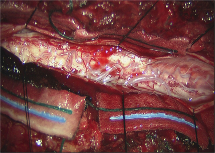
Case presentation: We present 10 cases of sdAVF treated at our center over an 8-year period. Seventy percent of patients were male. The mean age of presentation was 62.6 years. The most common lesion was a dorsal dural AVF with single feeder. All patients underwent open surgical ligation, six having preoperative coil embolization of the radicular artery to allow for intraoperative localization of the fistula. Eight patients showed improvement following treatment as graded by the Nurick system. Two patients failed to improve. None of the patients worsened. One patient had a radiation burn from the spinal angiogram requiring secondary closure and one patient had a pseudomeningocele at the site of surgery that resolved.
Discussion: The successful treatment of sdAVF requires a detailed understanding of clinical presentation and imaging findings to allow for precise treatment. Owing to the rarity of the condition, clinicians must continue to share their experiences to advance our knowledge.
Keywords: Spinal cord.
Conflict of interest statement
The authors declare no conflict of interest.
Figures


References
-
- Krings T, Lasjaunias PL, Hans FJ, Mull M, Nijenhuis RJ, Alvares H et al. Imaging in spinal vascular disease. Neuroimaging Clin N Am 2007; 17: 57–72. - PubMed
-
- Kendall BE, Logue V. Spinal epidural angiomatous malformations draining into intrathecal veins. Neuroradiology 1977; 13: 181–189. - PubMed
-
- Rashad S, Mohamed AB, Waseem A, Tamer H. Management of spinal dural arterio-venous fistulas. Report of 12 cases and review of literature. Clin Neurol Neurosurg 2014; 125: 81–86. - PubMed
-
- Merland JJ, Riche MC, Chiras J. Intraspinal extramedullary arteriovenous fistulae draining into the medullary veins. J Neuroradiol 1980; 7: 271–320. - PubMed
-
- Kim LJ, Spetzler RF. Classification and surgical management of spinal arteriovenous lesions: arteriovenous fistulae and arteriovenous malformations. Neurosurgery 2006; 59(Suppl 3): S195–S201. - PubMed
LinkOut - more resources
Full Text Sources
Other Literature Sources
