

Complete Atlantooccipital Assimilation with Basilar Invagination and Atlantoaxial Subluxation Treated Non-Surgically: A Case Report
- PMID: 28690960
- PMCID: PMC5501708
- DOI: 10.7759/cureus.1327
Complete Atlantooccipital Assimilation with Basilar Invagination and Atlantoaxial Subluxation Treated Non-Surgically: A Case Report
Abstract
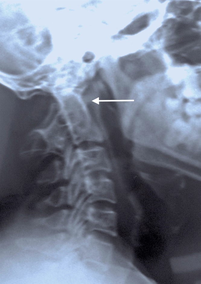
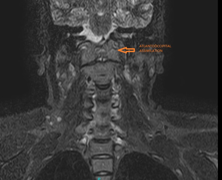
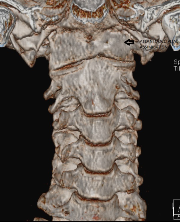
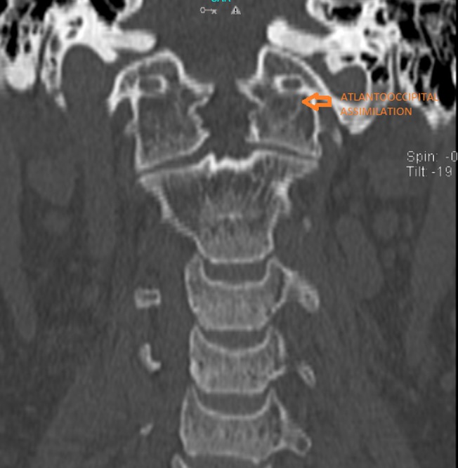
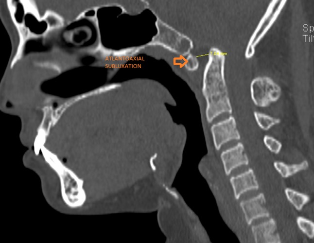
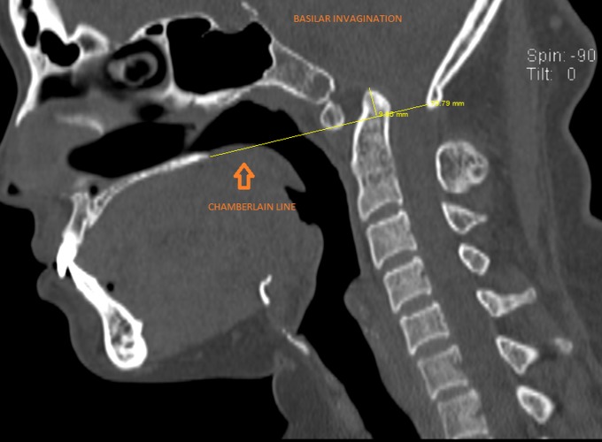
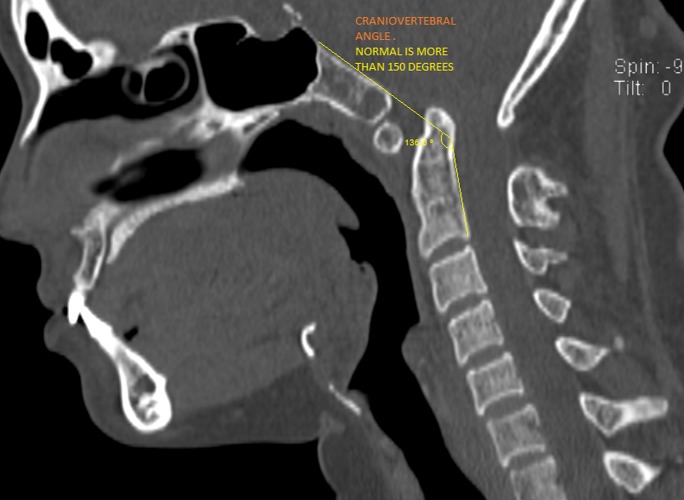
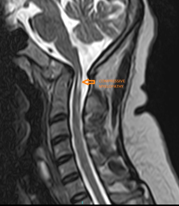
Atlantooccipital assimilation is a partial or complete congenital fusion between the atlas and the base of the occiput. Most patients with atlas assimilation are asymptomatic, but some may present with neurological problems such as myelopathy. We present the case of a 37-year-old woman who presented with neck and occipital pain, episodic neck stiffness, and dizziness. Medical imaging revealed complete atlantooccipital assimilation associated with basilar invagination, atlantoaxial subluxation, and predisposing anterior spinal cord compression. The patient was treated non-operatively with medications, cervical interferential therapy, and a rigid cervical orthosis.
Keywords: atlantoaxial subluxation; basilar invagination; complete atlantooccipital assimilation; predisposing anterior cord compression; treated non-surgically.
Conflict of interest statement
The authors have declared that no competing interests exist.
Figures

References
-
- Smoker WR, Khanna G. Childs Nerv Syst. Vol. 24. Oct: 2008. Imaging the craniocervical junction; pp. 1123–1145. - PubMed
-
- Congenital anomalies of the cervical spine. Hensinger RN. http://journals.lww.com/corr/Abstract/1991/03000/Congenital_Anomalies_of... Clin Orthop Relat Res. 1991;264:16–38. - PubMed
-
- Tsou PM, Yau A, Hodgson AR. Clin Orthop Relat Res. Vol. 152. Sep-Oct: 1980. Embryogenesis and prenatal development of congenital vertebral anomalies and their classification; pp. 211–231. - PubMed
-
- Muthukumar N, Swaminathan R, Venkatesh G, Bhanumathy SP. Acta Neurochir (Wien) Vol. 147. Aug-Sep: 2005. A morphometric analysis of the foramen magnum region as it relates to the transcondylar approach; pp. 889–895. - PubMed
-
- A case of foramen magnum syndrome caused by atlanto-occipital assimilation with intracanal fibrosis. Martellacci S, Ben Salem D, Méjean N, Sautreaux JL, Krausé D. Surg Radiol Anat. 2008;30:149–152. - PubMed
Publication types
LinkOut - more resources
Full Text Sources
Other Literature Sources