. 2017 Jun 22:4:87.
doi: 10.3389/fmed.2017.00087. eCollection 2017.

Fluorescence In Situ Hybridization for Diagnosis of Whipple's Disease in Formalin-Fixed Paraffin-Embedded Tissue

Affiliations

Fluorescence In Situ Hybridization for Diagnosis of Whipple's Disease in Formalin-Fixed Paraffin-Embedded Tissue

Peter Braubach et al. Front Med (Lausanne). .

Abstract

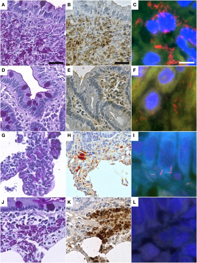
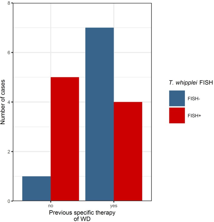
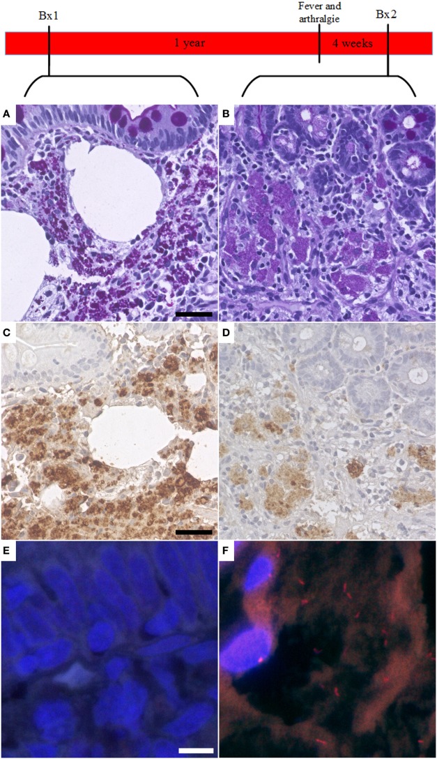
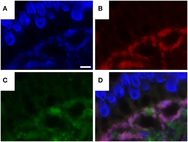
Whipple's disease (WD) is a rare chronic systemic infection with a wide range of clinical symptoms, routinely diagnosed in biopsies from the small intestine and other tissues by periodic acid-Schiff (PAS) diastase staining and immunohistological analysis with specific antibodies. The aim of our study was to improve the pathological diagnosis of WD. Therefore, we analyzed the potential of fluorescence in situ hybridization (FISH) for diagnosing WD, using a Tropheryma (T.) whipplei-specific probe. 19 formalin-fixed paraffin-embedded (FFPE) duodenal biopsy specimens of 12 patients with treated (6/12) and untreated (6/12) WD were retrospectively examined using PAS diastase staining, immunohistochemistry, and FISH. 20 biopsy specimens with normal intestinal mucosa, Helicobacter pylori, or mycobacterial infection, respectively, served as controls. We successfully detected T. whipplei in tissue biopsies with a sensitivity of 83% in untreated (5/6) and 40% in treated (4/10) cases of WD. In our study, we show that FISH-based diagnosis of individual vital T. whipplei in FFPE specimens is feasible and can be considered as ancillary diagnostic tool for the diagnosis of WD in FFPE material. We show that FISH not only detect active WD but also be helpful as an indicator for the efficiency of antibiotic treatment and for detection of recurrence of disease when the signal of PAS diastase and immunohistochemistry lags behind the recurrence of disease, especially if the clinical course of the patient and antimicrobial treatment is considered.

Keywords: Tropheryma whipplei; Whipple’s disease; fluorescence in situ hybridization; formalin-fixed paraffin-embedded tissue; immunohistochemistry.


Figures

Figure 1
Figure 1
Representative cases of Whipple’s disease with periodic acid-Schiff diastase staining (A,D,G,J) show accumulated macrophages intensely positive in periodic acid–Schiff diastase staining and with positive reaction in immunohistochemistry with a specific anti-Tropheryma whipplei (T. whipplei) antibody (B,E,H,K). Fluorescence in situ hybridization with a T. whipplei specific probe (C,F,I,L) shows differing numbers of intensely red-labeled bacteria ranging from dense aggregates (C) to sparse infiltrates of single bacteria (F,I), while some cases did not show bacteria in FISH (L). Presented cases are WD9 (A–C), WD1 (D–F), WD13 (G–I), WD17 (J–L) (see also Table 2). The black bar is 30 µm. The white bar is 5 µm.
Figure 2
Figure 2
Before specific antibiotic therapy, the majority of cases investigated in this series were positive in the Tropheryma whipplei (T. whipplei) specific fluorescence in situ hybridization (FISH). After specific therapy, the majority of cases were FISH-negative, remaining positivity may indicate persistence or recurrence of disease (see also Table 2).
Figure 3
Figure 3
Fluorescent in situ hybridization (FISH) of Tropheryma whipplei (T. whipplei) may be a marker of disease recurrence. In one case, the first available biopsy (Bx1, taken after specific therapy) showed PAS diastase positive macrophages in the intestinal mucosa (A) and was positive in immunohistochemistry with the specific T. whipplei antibody (C). However, it was negative in FISH (E). One year after the biopsy, the patient presented with fever and arthralgia. A second biopsy (Bx2) was taken 4 weeks after onset of symptoms. This biopsy showed a reduction in PAS diastase positivity (B) and intensity of the specific immunohistochemistry (D) consistent with an effective therapy; however, in this biopsy, vital T. whipplei organisms could be detected in FISH (F).
Figure 4
Figure 4
Combination of Tropheryma whipplei (T. whipplei) specific fluorescence in situ hybridization and fluorescence immunostaining with anti-T. whipplei antibody in an intestinal biopsy. 4′,6-Diamidin-2-phenylindol (DAPI) (A), T. whipplei-specific probe (Cy3) (B), anti- T. whipplei antibody with a fluorescence-labeled secondary antibody (Cy2) (C), overlay of 4′,6-DAPI, T. whipplei-specific probe (Cy3), and anti- T. whipplei antibody with a fluorescence-labeled secondary antibody (Cy2) (D). The white bar represents 5 µm.

References

    1. Marth T, Moos V, Müller C, Biagi F, Schneider T. Tropheryma whipplei infection and Whipple’s disease. Lancet Infect Dis (2016) 16:e13–22. 10.1016/S1473-3099(15)00537-X - DOI - PubMed
    1. Biagi F, Balduzzi D, Delvino P, Schiepatti A, Klersy C, Corazza GR. Prevalence of Whipple’s disease in north-western Italy. Eur J Clin Microbiol Infect Dis (2015) 34:1347–8. 10.1007/s10096-015-2357-2 - DOI - PubMed
    1. Relman DA, Schmidt TM, MacDermott RP, Stanley F. Identification of the uncultured Bacillus of Whipple’s disease. N Engl J Med (1992) 327:293–327. 10.1056/NEJM199207303270501 - DOI - PubMed
    1. Bonhomme CJ, Renesto P, Desnues B, Ghigo E, Lepidi H, Fourquet P, et al. Tropheryma whipplei glycosylation in the pathophysiologic profile of Whipple’s disease. J Infect Dis (2009) 199:1043–52. 10.1086/597277 - DOI - PubMed
    1. Fredricks DN, Relman DA. Localization of Tropheryma whipplei rRNA in tissues from patients with Whipple’s disease. J Infect Dis (2001) 183:1229–37. 10.1086/319684 - DOI - PubMed

LinkOut - more resources