Review
. 2018 Jun;24(3):261-276.
doi: 10.1177/1073858417717210. Epub 2017 Jul 8.

Plasticity in the Working Memory System: Life Span Changes and Response to Injury

Affiliations
Review

Plasticity in the Working Memory System: Life Span Changes and Response to Injury

Sean Froudist-Walsh et al. Neuroscientist. 2018 Jun.

Abstract

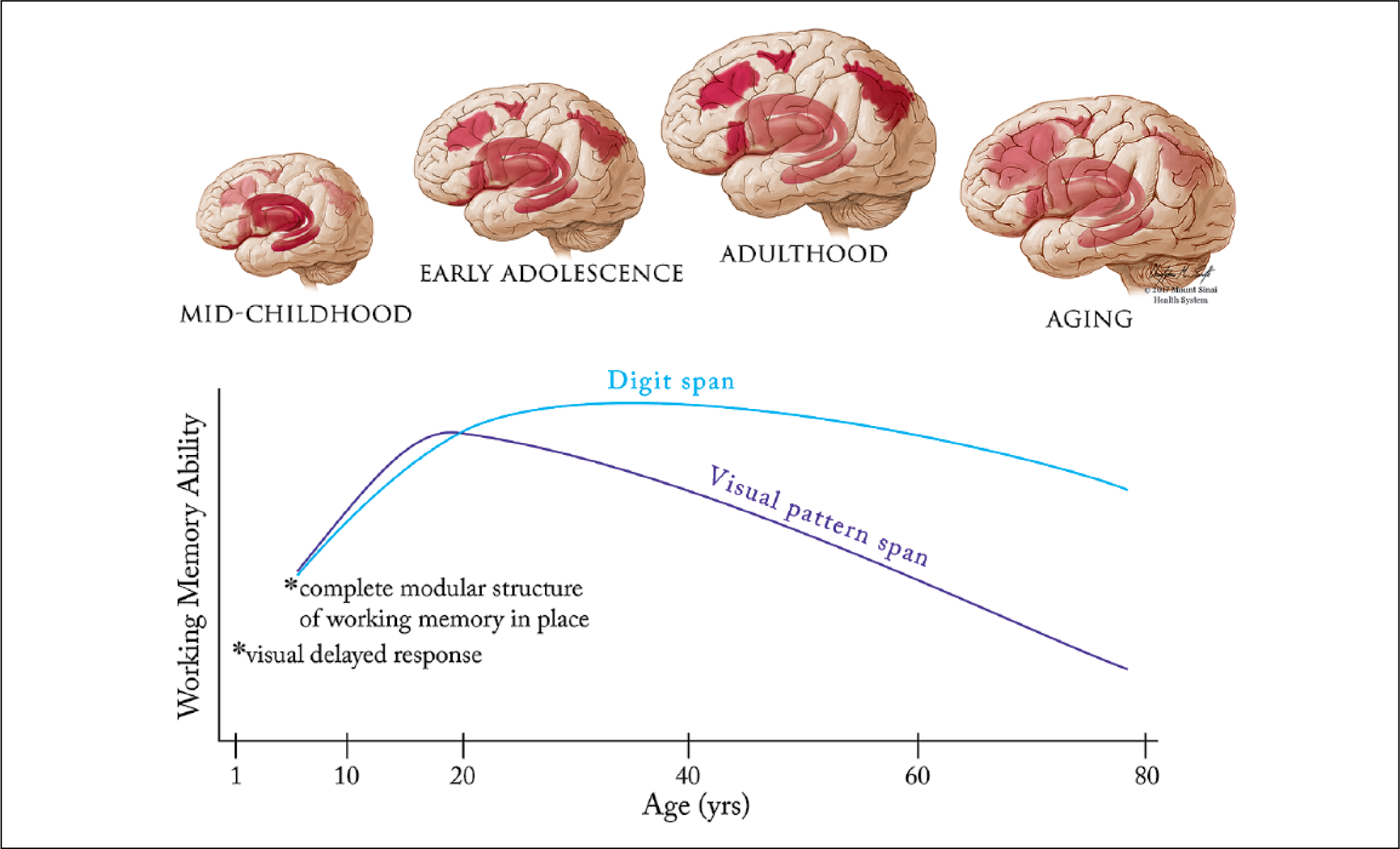
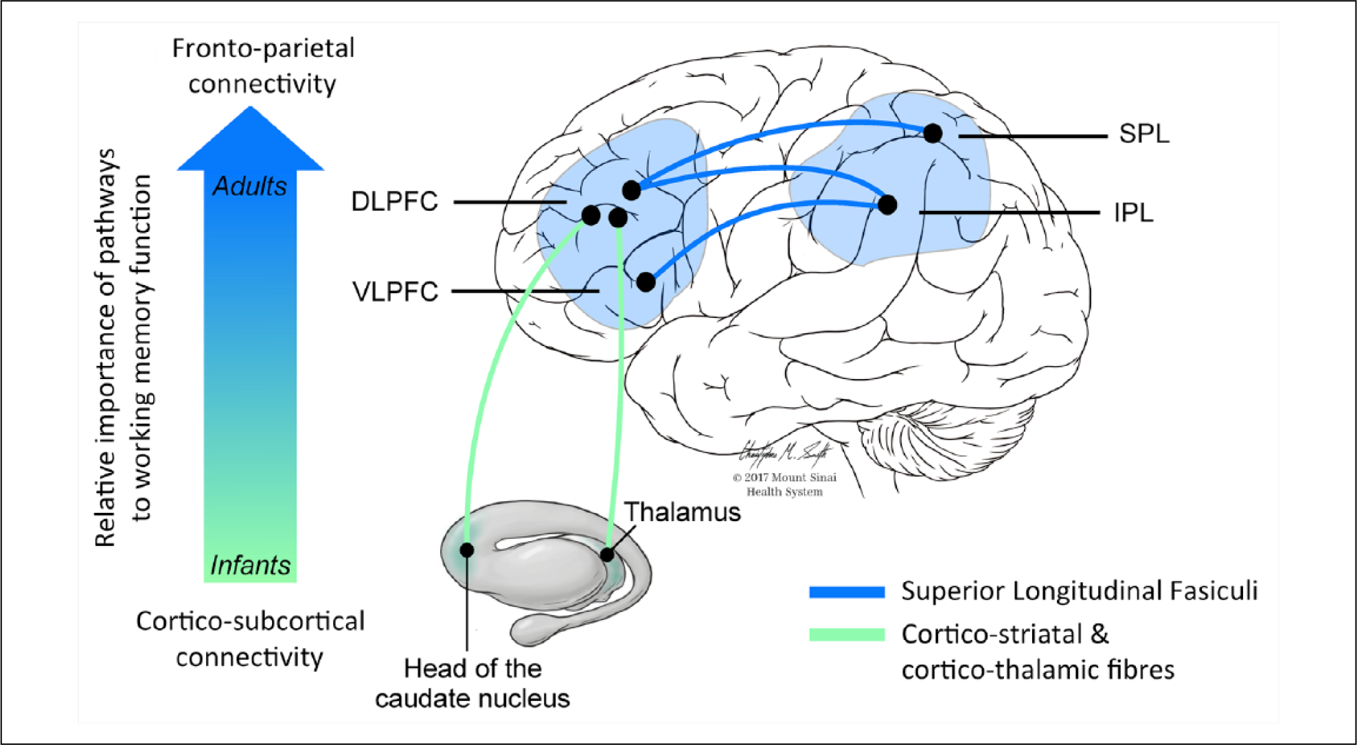
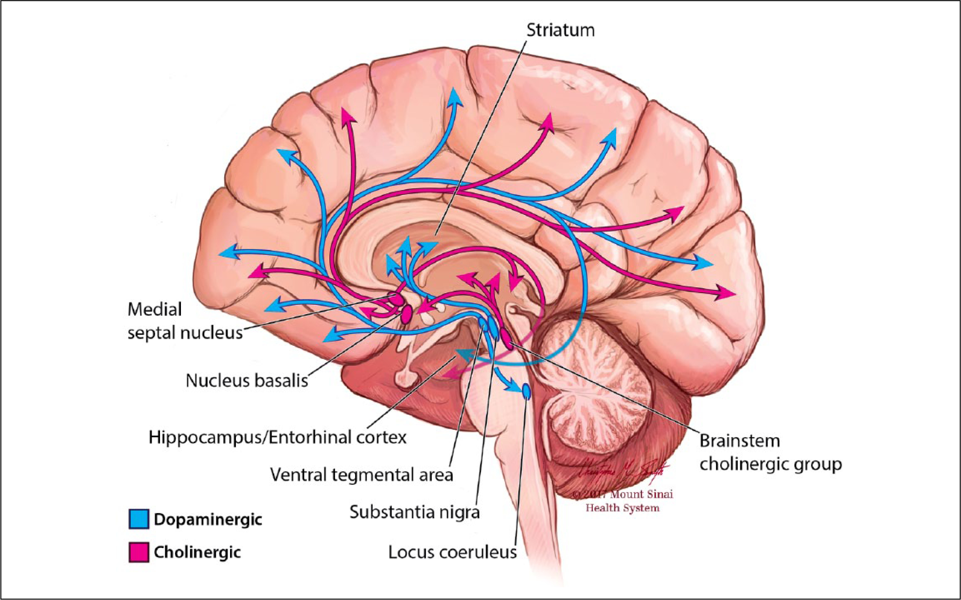
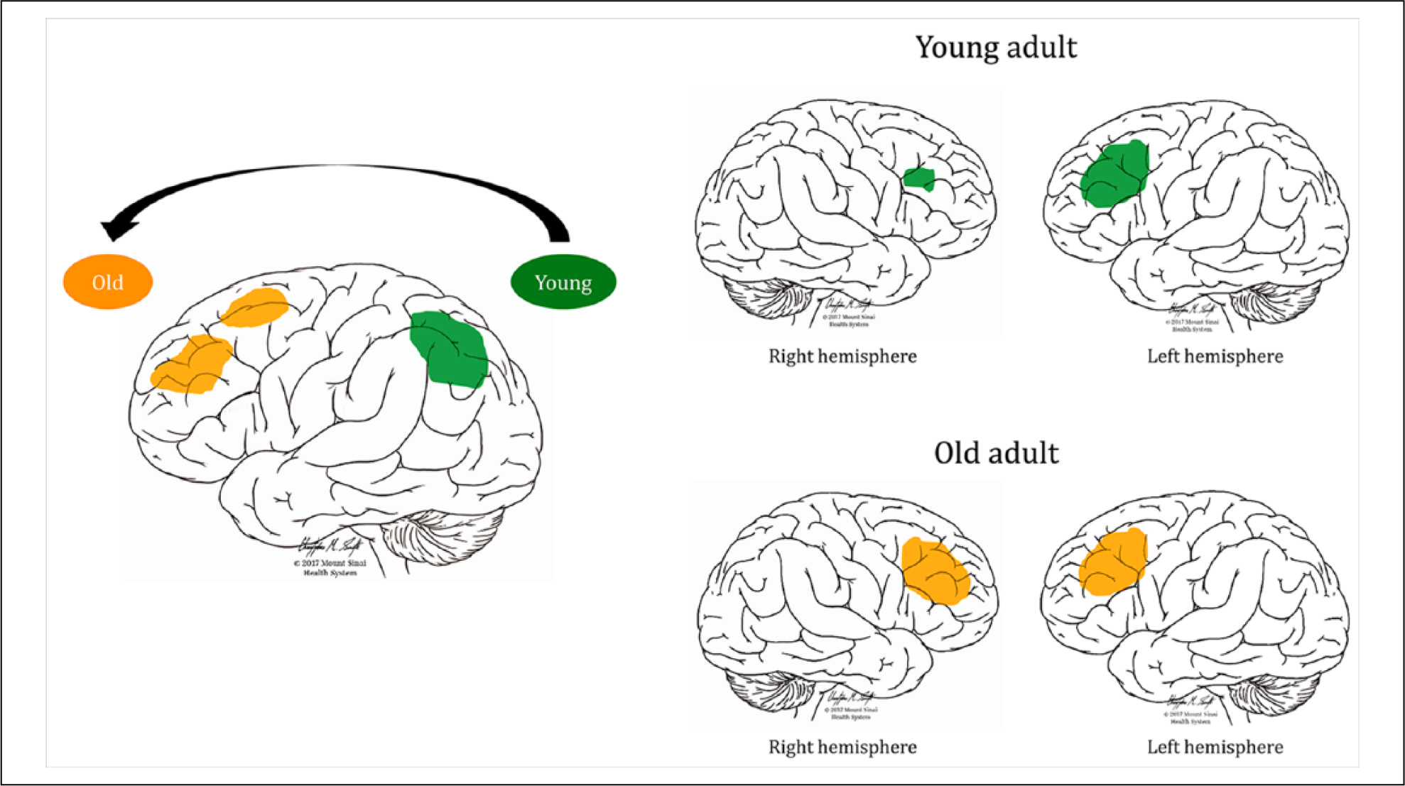
Working memory acts as a key bridge between perception, long-term memory, and action. The brain regions, connections, and neurotransmitters that underlie working memory undergo dramatic plastic changes during the life span, and in response to injury. Early life reliance on deep gray matter structures fades during adolescence as increasing reliance on prefrontal and parietal cortex accompanies the development of executive aspects of working memory. The rise and fall of working memory capacity and executive functions parallels the development and loss of neurotransmitter function in frontal cortical areas. Of the affected neurotransmitters, dopamine and acetylcholine modulate excitatory-inhibitory circuits that underlie working memory, are important for plasticity in the system, and are affected following preterm birth and adult brain injury. Pharmacological interventions to promote recovery of working memory abilities have had limited success, but hold promise if used in combination with behavioral training and brain stimulation. The intense study of working memory in a range of species, ages and following injuries has led to better understanding of the intrinsic plasticity mechanisms in the working memory system. The challenge now is to guide these mechanisms to better improve or restore working memory function.

Keywords: MRI; acetylcholine; aging; brain injury; dopamine; neurodevelopment; neurotransmitters; plasticity; preterm birth; working memory.


Conflict of interest statement

Declaration of Conflicting Interests

The author(s) declared the following potential conflicts of interest with respect to the research, authorship, and/or publication of this article: M.L.B. has received honoraria from Pfizer, Eisai, Janssen-España, Novartis, Lundbeck, and Nutricia and consultancy for fees from Merz, Eli Lilly, and GlaxoSmithKline. He has received speaking fees from Pfizer, Eisai, Janssen-España, Novartis, Lundbeck, and Nutricia. All other authors report no conflicts of interest.

Figures

Figure 1.
Figure 1.
Lifespan development of the working memory system. Top: Early in development working memory depends on subcortical and ventral cortical structures such as the striatum, thalamus, hippocampus and insula. After a transition phase in late childhood/early adolescence, working memory gradually begins to settle upon a network of frontoparietal cortical regions (dorsolateral and ventrolateral prefrontal cortex, frontal eye fields and posterior parietal cortex), centered on the dorsolateral prefrontal cortex. Finally, associations between cortical structures (in particular the parietal cortex) and working memory ability decline during aging. The reliance of working memory on specific structures at different stages in development is represented on an opacity scale (more opaque = greater involvement of the area in working memory). Bottom: Not all aspects of working memory show the same trajectory during development and aging. By one year of age, infants can perform basic visual delayed response tasks, while the complete modular structure (from Baddeley’s model – see Box 1) is in place by at least age 4. Adults at age 55 perform visual pattern span tasks worse than 8 year olds, but can perform digit span tasks on a par with young adults (Brockmole and Logie, 2013).
Figure 2.
Figure 2.
Schematic illustration of the white matter tracts mainly implicated in working memory processes at different points in the lifespan. The vertical arrow indicates how infants’ working memory functions rely initially on subcortical connections due to immature frontoparietal connections, while later in life there is a progressively greater involvement of the dorsolateral and ventrolateral prefrontal cortices connected to the parietal lobe through the three branches of the superior longitudinal fasciculus (SLF).
Figure 3.
Figure 3.
Dopamine and acetylcholine. Ascending projections of the dopaminergic and cholinergic systems from midbrain and basal forebrain innervate diverse cortical and subcortical targets.
Figure 4.
Figure 4.
Examples of working memory adaptations during healthy aging. The left image shows a posterior-anterior shift in activation during a complex visual selective attention task (Ansado and others 2012). Younger subjects show greater activation in the posterior parietal cortex bilaterally, whereas older subjects shift their activation to more frontal regions (Davis and others 2008; Ansado and others 2012). The right image shows the usual left-dominant asymmetric activation in young adults during a verbal working memory task (top) and more symmetrical bilateral activation of prefrontal brain regions in older adults during the same task to compensate for age-related neural deficits (bottom, Cabeza, 2002).
Figure 5.
Figure 5.
Different types of brain plasticity following lesions. Lesions or dysfunctions of a brain area alter the interaction between structurally normal regions and between normal and dysfunctional regions. (A) Interhemispheric transfer of a particular function from one lesioned area to its homotopic counterpart. (B) A normally functioning area can enlarge its volume in response to repetitive training or other treatments (drugs, brain stimulation). Dysfunctional unlesioned areas (as can occur during aging) can also show greater than normal activity during demanding tasks to in order maintain normal performance. (C) When an area is deprived from its main inputs (e.g. due to hearing loss, blindness, tract hypoplasia) patients can maintain normal task performance by receiving input from new areas. (D) New strategies using alternative spared regions and connections can be employed to undertake the affected task. Note that more than one type of plasticity (i.e., homologous area adaptation and ipsilateral map extension) can co-occur in a single subject. Green circles represent preserved brain areas, red circles fully lesioned/dysfunctional areas, and mixed red/green circles partially lesioned-dysfunctional areas. The blue circle represents vicarious compensation by an area not previously implicated in the function. Red lines represent interrupted connections, green lines normal connections, and dotted green lines new, alternative connections. See further information in Box 3 and text.

References

    1. Aanes S, Bjuland KJ, Skranes J, Løhaugen GCC. 2015. Memory function and hippocampal volumes in preterm born very-low-birth-weight (VLBW) young adults. Neuroimage 105:76–83. - PubMed
    1. Alcauter S, Lin W, Smith JK, Short SJ, Goldman BD, Reznick JS, and others. 2014. Development of thalamocortical connectivity during infancy and its cognitive correlations. J Neurosci 34:9067–75. - PMC - PubMed
    1. Alexander GE, Goldman PS. 1978. Functional development of the dorsolateral prefrontal cortex: an analysis utilizing reversible cryogenic depression. Brain Res 143:233–49. - PubMed
    1. Alloway TP, Alloway RG. 2013. Working memory across the lifespan: a cross-sectional approach. J Cogn Psychol 25:84–93.
    1. Alloway TP, Gathercole SE, Pickering SJ. 2006. Verbal and visuospatial short-term and working memory in children: are they separable? Child Dev 77:1698–716. - PubMed

Publication types

LinkOut - more resources