Review
. 2017 Jul 25;5(8):1421-1434.
doi: 10.1039/c7bm00295e.

Using biomaterials to rewire the process of wound repair

Affiliations
Review

Using biomaterials to rewire the process of wound repair

Anna Stejskalová et al. Biomater Sci. .

Abstract

Wound healing is one of the most complex processes that our bodies must perform. While our ability to repair wounds is often taken for granted, conditions such as diabetes, obesity, or simply old age can significantly impair this process. With the incidence of all three predicted to continue growing into the foreseeable future, there is an increasing push to develop strategies that facilitate healing. Biomaterials are an attractive approach for modulating all aspects of repair, and have the potential to steer the healing process towards regeneration. In this review, we will cover recent advances in developing biomaterials that actively modulate the process of wound healing, and will provide insight into how biomaterials can be used to simultaneously rewire multiple phases of the repair process.


Figures

Fig. 1
Fig. 1
Following haemostasis, the process of wound healing follows a progression of events including inflammation, generation of new tissue, and remodelling of the nascent tissue; these phases occur over timescales that range from hours to many months. Biomaterials can be used to augment the natural repair process at all stages of wound repair, depending on the aspect of the repair process that is looking to be modulated. Ideally, biomaterials are designed/chosen according to the underlying biological process that is being targeted – there is no determined set of biomaterials that are ideal for one aspect of the wound healing process. This diversity and complexity in options for biomaterials mirrors the complexity of the biology underlying the process of wound repair.
Fig. 2
Fig. 2
(A) PLPs circulate freely within blood due to no interactions with fibrinogen. (B) PLPs selectively bind fibrin protofibrils that begin to appear in the early stages of clot formation. (C) As the fibrin network grows, PLPs bond to multiple fibrin fibres. (D) The bonding interaction between PLPs and the fibrin network causes destabilisation and subsequent collapse of the network, in turn densifying the clot.
Fig. 3
Fig. 3
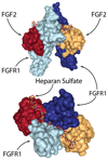
The FGF-2/FGFR1/heparan sulfate complex. Two FGF-2 molecules are tied together via a heparan sulfate chain that spans the middle of the FGFR1 dimer. Visualised from PDB 1FQ9 using NGL viewer.
Fig. 4
Fig. 4
Structure of a syndesome and alginate wound dressings containing syndesomes and FGF-2. (b) Full thickness skin wound healing in ob/ob mice on high fat diet. Open wound areas after 14 days for untreated, FGF-2 treated, syndesome treated (S4PL), and syndesome + FGF-2 treated mice. (c) Quantification of epidermal growth beyond edge of wound at day 14. Adapted with permission from ref. . Copyright 2016 John Wiley and Sons.
Fig. 5
Fig. 5
(A) LbL dressings assembled with VEGF-A165 and PDGF-BB lose distinct release profiles due to interdiffusion. (B) Layers of thiolated poly (acrylic acid) spontaneously form two-dimensional diffusion barriers within the film if spaced sufficiently far apart. The cross-linked layers serve as reversible diffusion barriers that enable individual control over the release kinetics of each growth factor. Adapted with permission from ref. . Copyright 2015 John Wiley and Sons.
Fig. 6
Fig. 6
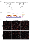
(a) Chemical structure of PEA and PMA. (b) HUVECs were seeded on surfaces coated with either PEA or PMA and fibronectin (FN), and then covered with a thin layer of fibrin. (c) Fluorescence images of HUVECs after 6 days culture on either FN surfaces, VEGF coated FN surfaces (VEGFc), or FN coated surfaces with VEGF in the media (VEGFm). Adapted from ref. under permission of a creative commons license.

References

    1. Robbins JM, Strauss G, Aron D, Long J, Kuba J, Kaplan Y. J Am Podiatr Med Assoc. 2008;98:489–493. - PubMed
    1. Shah JB. J Am Col Certif Wound Spec. 2011;3:65–66. - PMC - PubMed
    1. Broughton G, Janis JE, Attinger CE. Plast Reconstr Surg. 2006;117:6S–11S. - PubMed
    1. Philip R. Can Med Assoc J. 1929;21:650–655. - PMC - PubMed
    1. Finnegan S, Percival SL. Adv Wound Care. 2015;4:398–406. - PMC - PubMed

MeSH terms

Substances