. 2017 Sep;15(9):1867-1877.
doi: 10.1111/jth.13775. Epub 2017 Aug 9.

A discontinuous autoinhibitory module masks the A1 domain of von Willebrand factor

Affiliations

A discontinuous autoinhibitory module masks the A1 domain of von Willebrand factor

W Deng et al. J Thromb Haemost. 2017 Sep.

Abstract

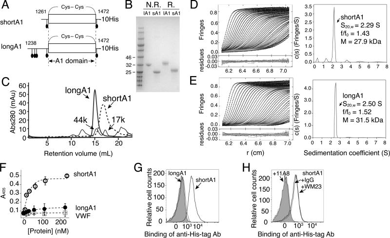
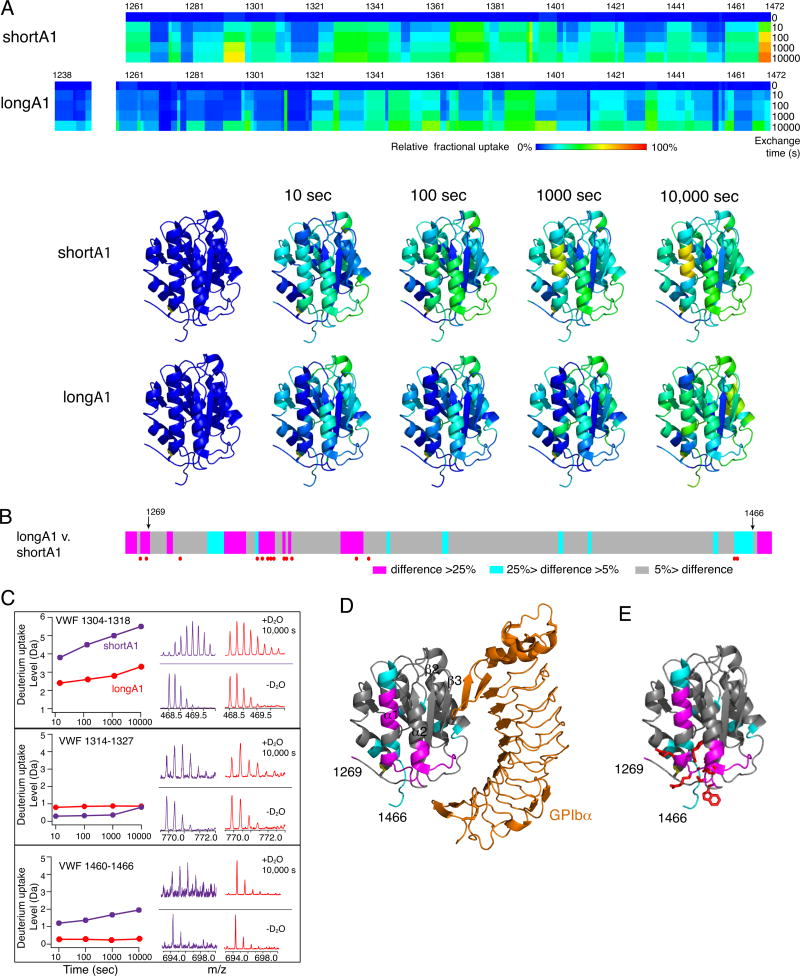
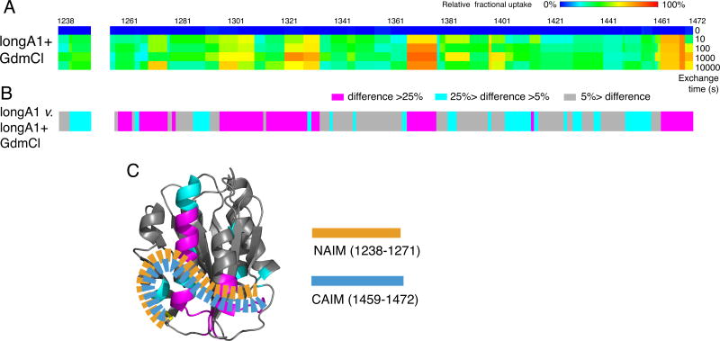
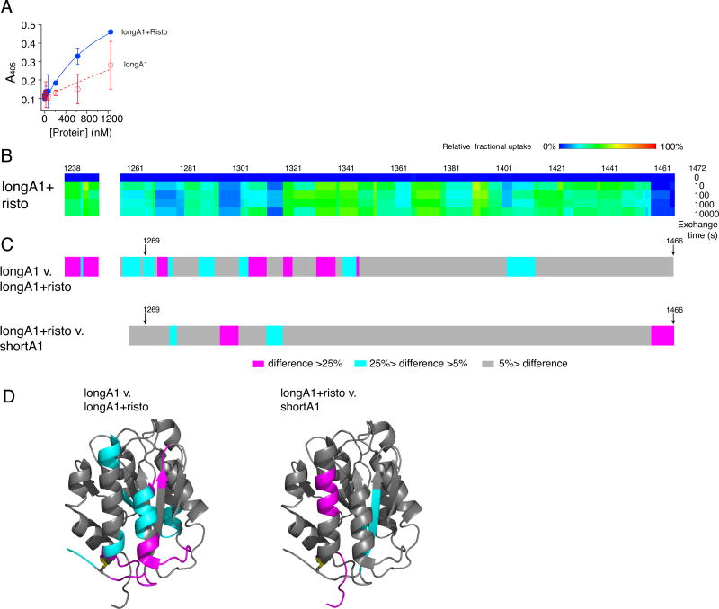
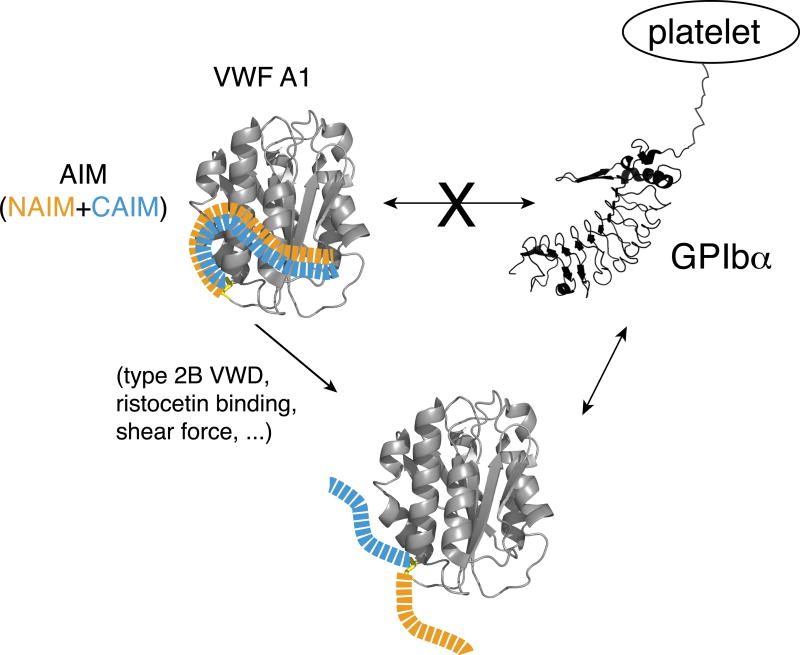
Essentials The mechanism for the auto-inhibition of von Willebrand factor (VWF) remains unclear. Hydrogen exchange of two VWF A1 fragments with disparate activities was measured and compared. Discontinuous residues flanking A1 form a structural module that blocks A1 binding to the platelet. Our results suggest a potentially unified model of VWF activation. Click to hear an ISTH Academy presentation on the domain architecture of VWF and activation by elongational flow by Dr Springer SUMMARY: Background How von Willebrand factor (VWF) senses and responds to shear flow remains unclear. In the absence of shear flow, VWF or its fragments can be induced to bind spontaneously to platelet GPIbα. Objectives To elucidate the auto-inhibition mechanism of VWF. Methods Hydrogen-deuterium exchange (HDX) of two recombinant VWF fragments expressed from baby hamster kidney cells were measured and compared. Results The shortA1 protein contains VWF residues 1261-1472 and binds GPIbα with a significantly higher affinity than the longA1 protein that contains VWF residues 1238-1472. Both proteins contain the VWF A1 domain (residues 1272-1458). Many residues in longA1, particularly those in the N- and C-terminal sequences flanking the A1 domain, and in helix α1, loops α1β2 and β3α2, demonstrated markedly reduced HDX compared with their counterparts in shortA1. The HDX-protected region in longA1 overlaps with the GPIbα-binding interface and is clustered with type 2B von Willebrand disease (VWD) mutations. Additional comparison with the HDX of denatured longA1 and ristocetin-bound longA1 indicates the N- and C-terminal sequences flanking the A1 domain form cooperatively an integrated autoinhibitory module (AIM) that interacts with the HDX-protected region. Binding of ristocetin to the C-terminal part of the AIM desorbs the AIM from A1 and enables longA1 binding to GPIbα. Conclusion The discontinuous AIM binds the A1 domain and prevents it from binding to GPIbα, which has significant implications for the pathogenesis of type 2B VWD and the shear-induced activation of VWF activity.

Keywords: Type 2B von Willebrand disease; blood platelet; ristocetin; tandem mass spectrometry; von Willebrand factor.


Conflict of interest statement

Disclosure of Conflict of Interests

The authors state that they have no conflict of interest.

Figures

Figure 1
Figure 1. Recombinant shortA1 and longA1 proteins are primarily monomeric, and only ShortA1, but not longA1, binds GPIbα
(A) Illustrations of shortA1 and longA1 proteins. Both proteins contain a C-terminal decahistidine tag. Corresponding VWF residues in each protein are marked by the starting and ending residue numbers. O-glycosylation sites (filled ovals) and the 1272–1458 disulfide bond delimiting the A1 domain are marked. (B) SDS-PAGE of purified longA1 (lA1) and shortA1 (sA1) under non-reducing (N.R.) and reducing (R.) conditions. The gel was stained by Coomassie blue. Nearby molecular weight markers are marked on the left. (C) Superimposed gel filtration chromatograms of shortA1, longA1 and molecular weight standards as indicated by arrows. (D, E) Sedimentation velocity results for shortA1 (D) and longA1 (E) showing fitted interference scans and residuals (left) and sedimentation coefficient distributions (right). Only every 2nd scan and every 3rd data points are shown for clarity. (F) Isotherms of shortA1 (open circle), longA1 (filled circle) and human VWF (open squares) binding to immobilized GPIb-IX measured by ELISA (n=3). (G) Flow cytometry histograms showing binding of shortA1 and lack of binding of longA1 to human platelets. Gray: platelets incubated with only FITC-labeled irrelevant IgG. (H) Overlaid histograms showing that binding of shortA1 to human platelets is inhibited by anti-GPIbα ligand-binding domain MAb 11A8 but not by anti-GPIbα macroglycopeptide region MAb WM23.
Figure 2
Figure 2. Difference in HDX between shortA1 and longA1
Comparison of HDX between shortA1 and longA1. (A) Residual HDX heat maps of shortA1 and longA1 for the noted exchange time. The line for 0 sec denotes the results obtained without exchange. Relative fractional deuterium uptake was calculated for each residue amide from the measured deuterium uptakes of peptic fragments as described in Materials and Methods, and plotted using the rainbow color scale in the figure. Residues 1251–1257 were not detected and are left blank. HDX data of shortA1 and longA1 at the indicated exchange time are mapped to the structure of A1 domain (PDB: 1SQ0; only residues 1269–1466 are shown in the structure). The ribbon diagram of the complex structure is generated using PyMOL. (B) The difference in relative fractional uptake between the same residues of longA1 and shortA1 after 10,000 s of exchange, as defined by the color code in the figure. Residues with reported type 2B VWD mutations [13] are marked by red dots. Residues 1269 and 1466, starting and ending residues of the structure of A1, respectively, are marked by arrows. (C) Representative plots of HDX and mass spectra of three peptic fragments from shortA1 and longA1. Each peptide is identified by the starting and ending residue numbers of VWF. (D) The difference in HDX mapped to a structure of A1 in complex with the ligand-binding domain of GPIbα. Certain secondary structure elements in A1 are labeled. (E) The HDX-protected region in the A1 domain (residues 1269–1466) is clustered with type 2B VWD mutations. Each mutation site is illustrated by its side chain shown in red sticks.
Figure 3
Figure 3. AIM consists of two discontinuous segments flanking the A1 domain
(A) Residual HDX heat maps of longA1 in 4 M GdmCl (longA1+GdmCl) for the noted exchange time, following the same format as described in Figure 3A. (B) The difference bars following the same format as Figure 3B. (C) Illustration of the AIM masking the HDX-protected region in the A1 domain of longA1. AIM is shown as orange and celestial blue dashed ribbons, indicative of well-integrated NAIM and CAIM sequences.
Figure 4
Figure 4. Addition of ristocetin desorbs the AIM from A1 and facilitates longA1 binding to GPIbα
(A) Binding isotherms of longA1 in the presence (blue) and absence (red) of 1.5 mg/mL ristocetin. (B) Residual HDX heat maps of longA1 mixed with ristocetin (longA1+risto) for the noted exchange time, following the same format as Figure 3A. Residues 1252–1258 and 1467–1472 were not detected and are left blank. (C) The plots of difference in the relative fractional uptake between the same residues of longA1 and longA1+risto, and between longA1+risto and shortA1, following the same format as Figure 3B. (D) The differences are mapped to structures of the A1 domain (PDB: 1SQ0).
Figure 5
Figure 5. Model of the AIM-masking of A1
A model of AIM masking of the A1 domain in VWF. In VWF, the N- and C-terminal sequences flanking the A1 domain, designated as NAIM (orange) and CAIM (celestial blue), respectively, cooperatively form the AIM that binds the A1 domain and blocks its association with GPIbα. Various factors, including ristocetin, type 2B VWD mutations and shear force, may induce VWF binding to GPIbα by destabilizing the AIM/A1 association and/or disrupting AIM.

References

    1. Wagner DD. Cell biology of von Willebrand factor. Annu Rev Cell Biol. 1990;6:217–246. - PubMed
    1. Sadler JE. Biochemistry and genetics of von Willebrand factor. Annu Rev Biochem. 1998;67:395–424. - PubMed
    1. Slayter H, Loscalzo J, Bockenstedt P, Handin RI. Native conformation of human von Willebrand protein. Analysis by electron microscopy and quasi-elastic light scattering. J. Biol.Chem. 1985;260:8559–8563. - PubMed
    1. Dong JF, Moake JL, Nolasco L, Bernardo A, Arceneaux W, Shrimpton CN, Schade AJ, McIntire LV, Fujikawa K, Lopez JA. ADAMTS-13 rapidly cleaves newly secreted ultralarge von Willebrand factor multimers on the endothelial surface under flowing conditions. Blood. 2002;100:4033–4039. - PubMed
    1. Siedlecki CA, Lestini BJ, Kottke-Marchant KK, Eppell SJ, Wilson DL, Marchant RE. Shear-dependent changes in the three-dimensional structure of human von Willebrand factor. Blood. 1996;88:2939–2950. - PubMed

Publication types

MeSH terms