. 2017 Sep 15;12(9):2313-2323.
doi: 10.1021/acschembio.7b00361. Epub 2017 Aug 1.

Site-Specific Phosphorylation of PSD-95 PDZ Domains Reveals Fine-Tuned Regulation of Protein-Protein Interactions

Affiliations

Site-Specific Phosphorylation of PSD-95 PDZ Domains Reveals Fine-Tuned Regulation of Protein-Protein Interactions

Søren W Pedersen et al. ACS Chem Biol. .

Abstract

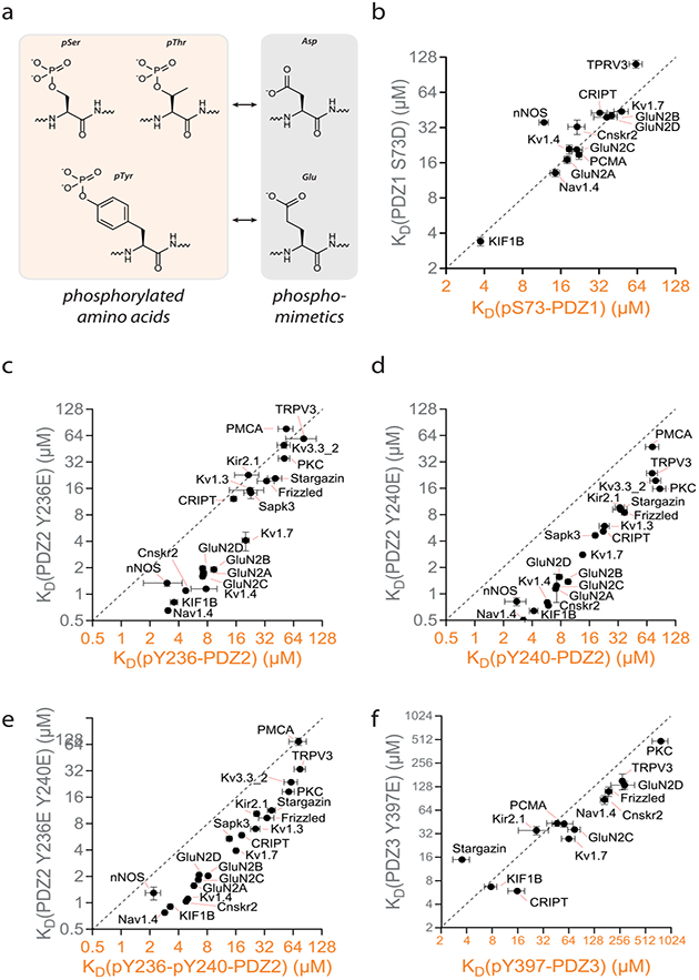
The postsynaptic density protein of 95 kDa (PSD-95) is a key scaffolding protein that controls signaling at synapses in the brain through interactions of its PDZ domains with the C-termini of receptors, ion channels, and enzymes. PSD-95 is highly regulated by phosphorylation. To explore the effect of phosphorylation on PSD-95, we used semisynthetic strategies to introduce phosphorylated amino acids at four positions within the PDZ domains and examined the effects on interactions with a large set of binding partners. We observed complex effects on affinity. Most notably, phosphorylation at Y397 induced a significant increase in affinity for stargazin, as confirmed by NMR and single molecule FRET. Additionally, we compared the effects of phosphorylation to phosphomimetic mutations, which revealed that phosphomimetics are ineffective substitutes for tyrosine phosphorylation. Our strategy to generate site-specifically phosphorylated PDZ domains provides a detailed understanding of the role of phosphorylation in the regulation of PSD-95 interactions.


Conflict of interest statement

The authors declare no competing financial interest.

Figures

Figure 1.
Figure 1.
Phosphorylation sites of PSD-95. (a) Organization of the five domains in PSD-95: PDZ1−3, an SH3 domain, and a GK domain. (b) Primary sequence of the human PSD-95 with phosphorylation sites identified though MS-MS studies (orange) or prediction tools (yellow).
Figure 2.
Figure 2.
Semisynthesis of phosphorylated PDZ1, PDZ2, and PDZ3 of PSD-95. (a) Semisynthetic strategy for obtaining N-terminal phosphorylation of PDZ1. The N-terminal fragment (ΔNPDZ1, 23 amino acids) was synthesized on 2-chlorotrityl (2CL) resin treated with hydrazine and subsequent chain elongation by SPPS generating a peptide containing a benzylated serine phosphate at position 73, which after trifluoroacetic acid (TFA) treatment cleaved the deprotected hydrazide phospho-peptide from the solid support. The C-terminal fragment (ΔCPDZ1, 69 AA) was recombinantly expressed in E. coli as a polyhistidine fusion protein containing a factor Xa (FXa) recognition site (IEGR) generating a N-terminal Cys after treatment with FXa enzyme. Ligation was initiated by oxidizing hydrazine of ΔNPDZ1 with NaNO2 for in situ generation of a C-terminal azide, which subsequently underwent thiolysis forming a thioester by the addition of 4-mercaptophenylacetic acid (MPAA). Ligation was performed by adding ΔCPDZ1 generating the full-length PDZ1 domain (Table S3). (b) Semisynthetic strategy for obtaining C-terminal phosphorylated PDZ2 and PDZ3. The C-terminal phosphorylations were introduced by synthesis of a N-terminal Cys peptide fragment (19 AA for ΔCPDZ2 and 26 AA for ΔCPDZ3) containing one or two phosphorodiamidate-protected tyrosines yielding pTyr after TFA treatment. The larger N-terminal fragment (ΔNPDZ2 and ΔNPDZ3 of both 75 AA) was produced through recombinant expression of a fusion protein containing the desired PDZ fragment, a Mycobacterium xenopi Gyrase A (Mxe GyrA) intein and a polyhistidine tag. Adding 2-mercaptoethanesulfonate (MESNa) induced a thiolysis reaction, leaving the extein fragment with a C-terminal thioester. The C- and N-terminal fragment were ligated followed by a desulfurization step converting Cys to Ala. The full-length semisynthetic PDZ domains were refolded by diafiltration. (c) The three PDZ domains with the four phosphorylation sites are displayed. Structures are adapted from X-ray crystal structures of PDZ12 (PDB-ID 3ZRT) and PDZ3 (PDB-ID 1BE9). (d) LC-MS traces for the five purified semisynthetic PDZ domains. Inlet shows the deconvoluted mass (expected masses: pS73-PDZ1, 9910.5 Da; PDZ2 pY236 and pY240/pY236-pY240, 10635/10715 Da; and PDZ3 pY397, 10958 Da).
Figure 3.
Figure 3.
Change in canonical ligand binding properties of phosphorylated PDZ domains. The fold change affinity toward C-terminal peptide ligands induced by single and double phosphorylation. Affinity increase is displayed as a left shifted fold change, whereas affinity decrease is displayed as a right shifted fold change. Fold changes for ligands having KD > 100 μM for nonphosphorylated proteins are not displayed.
Figure 4.
Figure 4.
Molecular details of the PDZ3−stargazin interaction. Model displaying the predicted interactions with side chains of two arginines of the stargazin C-terminal peptide and the Y397 phosphorylation of PDZ3. The R318 makes a salt bridge with the negative charge of pY397 (top left inlet). The positive charged R319 is stabilized by salt bridges originating from two positive charges of E331 and E364 of PDZ3 (bottom left inset). 3D model designed from the PDZ3 X-ray crystal structure of SAP-97 cocrystallized with the pentameric peptide (RTTPV) resembling C-terminal stargazin (PDB-ID: RJXT).
Figure 5.
Figure 5.
Structural homogeneity and stargazin binding investigated by the 1D 1H NMR spectrum of PDZ3 WT and PDZ3 pY397. (a) The well-dispersed spectra and overall similarity of the spectra indicate that PDZ3 WT and PDZ3 pY397 are both well folded and the phosphorylation does not alter the overall conformation of the PDZ domain. (b) A selected methyl peak of the 1H NMR spectra showing that the free and the stargazin peptide-bound forms of PDZ3 WT undergo fast to intermediate exchange, whereas the free and the stargazin peptide-bound forms of PDZ3 pY397 undergo slow exchange.
Figure 6.
Figure 6.
Kinetics of PDZ3 and stargazin interaction studied by single molecule FRET. (a) Cartoon representation of the single molecule FRET assay used to directly observe binding interactions between PDZ3 and stargazin. The full cytoplasmic domain of stargazin was labeled with the FRET acceptor (blue star) and attached to a passivated streptavidin surface via N-terminal biotinylation. PDZ3 was labeled with the FRET donor (magenta star) and added in solution at 100 nM. TIRF microscopy allows selective excitation of molecules at the surface. PDZ3 binding to stargazin brings the donor and acceptor close enough together to produce FRET. (b) Representative single molecule time trace of acceptor intensity. The bar above the panels illustrates the pattern of alternating laser excitation. First, the acceptor is directly excited to identify stargazin molecules on the surface. Next, the donor is excited, and individual protein interactions are visible as bursts of acceptor intensity produced by FRET. Finally, the acceptor is directly excited to probe for acceptor photobleaching, which has not occurred in these traces. (c) Histograms of the dwell times in the bound state (top) and unbound state (bottom) for the interaction between PDZ3 and stargazin. Data are shown as circles for PDZ3 WT (black), Y397E (gray), and pY397 (orange). Data from replicate measurements were pooled and fit as a single histogram (solid lines).
Figure 7.
Figure 7.
Comparing phosphorylations and phosphomimetic approaches for PDZ domains. (a) Structural differences of phosphorylated amino acids (orange background) and phosphomimetics (gray background). The KD values obtained from phosphorylated PDZ domains (x-axis, orange) and PDZ domains containing phosphomimetic mutations (y-axis, gray) compared for (b) PDZ1 S73, (c) PDZ2 Y236, (d) PDZ2 Y240, (e) PDZ2 Y236 Y240, and (f) PDZ3 Y397. Dashed lines: phosphomimics induce the same effect on ligand binding as the corresponding phosphor amino acids (KD(pS or pY) = KD(D or E)).

References

    1. Sheng M, and Sala C (2001) PDZ domains and the organization of supramolecular complexes. Annu. Rev. Neurosci 24, 1–29. - PubMed
    1. Kornau HC, Schenker LT, Kennedy MB, and Seeburg PH (1995) Domain interaction between NMDA receptor subunits and the postsynaptic density protein PSD-95. Science 269, 1737–1740. - PubMed
    1. Feng W, and Zhang M (2009) Organization and dynamics of PDZ-domain-related supramodules in the postsynaptic density. Nat. Rev. Neurosci 10, 87–99. - PubMed
    1. Zheng CY, Seabold GK, Horak M, and Petralia RS (2011) MAGUKs, synaptic development, and synaptic plasticity. Neuroscientist 17, 493–512. - PMC - PubMed
    1. Oliva C, Escobedo P, Astorga C, Molina C, and Sierralta J (2012) Role of the MAGUK protein family in synapse formation and function. Dev. Neurobiol 72, 57–72. - PubMed

Publication types

MeSH terms