. 2017 Aug;65(8):431-444.
doi: 10.1369/0022155417719419. Epub 2017 Jul 10.

Multiplex Staining by Sequential Immunostaining and Antibody Removal on Routine Tissue Sections

Affiliations

Multiplex Staining by Sequential Immunostaining and Antibody Removal on Routine Tissue Sections

Maddalena Maria Bolognesi et al. J Histochem Cytochem. 2017 Aug.

Abstract

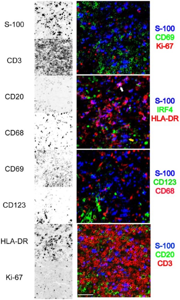
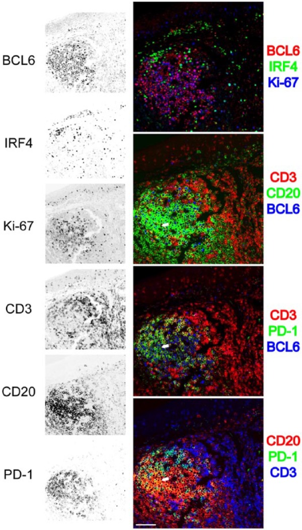
Multiplexing, labeling for multiple immunostains in the very same cell or tissue section in situ, has raised considerable interest. The methods proposed include the use of labeled primary antibodies, spectral separation of fluorochromes, bleaching of the fluorophores or chromogens, blocking of previous antibody layers, all in various combinations. The major obstacles to the diffusion of this technique are high costs in custom antibodies and instruments, low throughput, and scarcity of specialized skills or facilities. We have validated a method based on common primary and secondary antibodies and diffusely available fluorescent image scanners. It entails rounds of four-color indirect immunofluorescence, image acquisition, and removal (stripping) of the antibodies, before another stain is applied. The images are digitally registered and the autofluorescence is subtracted. Removal of antibodies is accomplished by disulfide cleavage and a detergent or by a chaotropic salt treatment, this latter followed by antigen refolding. More than 30 different antibody stains can be applied to one single section from routinely fixed and embedded tissue. This method requires a modest investment in hardware and materials and uses freeware image analysis software. Multiplexing on routine tissue sections is a high throughput tool for in situ characterization of neoplastic, reactive, inflammatory, and normal cells.

Keywords: antibody removal; epitope; immunofluorescence; multiplex.


Conflict of interest statement

Competing Interests: The author(s) declared no potential conflicts of interest with respect to the research, authorship, and/or publication of this article.

Figures

Figure 1.
Figure 1.
Variability for nine markers over 10 staining and stripping 2ME/SDS cycles. One single section for every three markers was stained (time 0) and sequentially stripped and restained for the same markers 10 times. Variation in staining intensity is expressed as a fraction of the 256 8bit channels over the initial staining intensity. Primary Ab incubation time: 1 hr, 2nd Ab: 30 min, single indirect IF. Data are from four independent 10-cycle experiments. Abbreviations: 2ME/SDS, beta-mercaptoethanol/sodium dodecyl sulfate; Ab = antibody; IF, immunofluorescence.
Figure 2.
Figure 2.
Comparison of antibody removal efficiency of 2ME/SDS and GnHCl. Tonsil sections were incubated with a FITC-conjugated anti-human IgM, stripped with either 2ME/SDS or GnHCl, counterstained with an AP-conjugated anti-FITC antibody and developed in NBT-BCIP. 2ME/SDS stripping (middle) leaves no stainable primary antibody. Stripping by GnHCl (bottom) leaves IgM+ plasma cells. Identical results are obtained with an anti-Rabbit AP-conjugated (not shown). High magnification in the insets. Scale bar: 500 µm. Abbreviations: 2ME/SDS, beta-mercaptoethanol/sodium dodecyl sulfate; GnHCl, guanidinium hydrochloride; FITC, Fluorescein Isothiocyanate; AP, alkaline phosphatase; NBT-BCIP, 5-bromo-4-chloro-3′-indolyl phosphate.
Figure 3.
Figure 3.
Variability for nine markers over 10 staining and stripping GnHCl, 6-M urea cycles. One single section for every three markers was stained (time 0) and sequentially stripped and restained for the same markers 10 times. Variation in staining intensity is expressed as a fraction of the 256 8bit channels over the initial staining intensity. Primary Ab incubation time: 1 hr, 2nd Ab: 30 min, single indirect IF. Abbreviations: Ab = antibody; IF, immunofluorescence.
Figure 4.
Figure 4.
Effect of NaBH4 15 mM alone or in combination with SDS on bound primary antibodies. Stripping is enhanced by SDS and the effect is proportional to the starting abundance of the target. Bcl-2, mouse IgG1; CD20, mouse IgG2a; CD3, rabbit Ig; IRF4, goat Ig; Ker8+19, pooled rabbit Ig anti-keratin 8 and 19. Abbreviations: NaBH4, sodium borohydride; SDS, sodium dodecyl phosphate.
Figure 5.
Figure 5.
Effect of NaBH4 0.6, 6, and 15 mM on antigens. Sections have been treated with 10 cycles of 10 min NaBH4 pH 9, followed by pH 4 buffer. Channel intensity variation over 256 channels is shown. Abbreviation: NaBH4, sodium borohydride.
Figure 6.
Figure 6.
Effect of stripping and restaining on abundant antigens. Sections in duplicate were stained in double indirect IF for the indicated abundant antigens (intermediate filaments). The channel value (A) or the % changes from the time 0 stain over 256 channels (B) ± SD are plotted before (Stain), after stripping (Strip), and after restaining with species- and isotype-matched nonimmune control Abs (Restain). Primary Ab incubation time: O/N, 2nd Ab: 30 min, double indirect IF. Abbreviations: Ab = antibody; IF, immunofluorescence.
Figure 7.
Figure 7.
Cycle-dependent decrease of kidney tissue AF. Variation over time of the kidney tubules AF (em 520 nm), expressed as % channel changes from time 0, over 10 staining and 2ME/SDS stripping cycles. Eleven different areas from three samples have been imaged, and the average intensity change over the starting AF is expressed as % change over 256 channels ± SD. Abbreviations: AF, autofluorescence; 2ME/SDS, beta-mercaptoethanol/sodium dodecyl sulfate; SD, standard deviation.
Figure 8.
Figure 8.
Detail of a multiplexed interfollicular tonsil area. Eight stains out of a 32-antibody multiplex are selected from a tonsil interfollicular area and shown as single stains (inverted gray scale) on the left, or combined into four three-color RGB composites. The area contains numerous S-100+ dendritic cells, HLA-DR+ with exceptions (yellow arrow), occasionally IRF4+ (white arrow), negative for Ki-67, CD69, CD68, and CD123. The dendritic cells are located in a CD3+ T-cell area, without contact with CD20+ B cells. GnHCl stripping method. Scale bar = 100 µm. Abbreviations: RGB, red green blue additive color; GnHCl, guanidinium hydrochloride.
Figure 9.
Figure 9.
Detail of a multiplexed tonsil follicle. Six stains out of a 32-antibody multiplex are selected from a tonsil follicle and shown as single stains (inverted gray scale) on the left, or combined into four three-color RGB composites. Proliferating (Ki-67+) germinal center BCL6+ CD20+ B cells are mutually exclusive stained for IRF4. PD-1 stains CD3+ T cells in the germinal center as well as rare PD-1+, BCL6+, CD20+ centroblasts (white arrow). GnHCl stripping method. Scale bar = 100 µm. Abbreviations: RGB, red green blue additive color; GnHCl, guanidinium hydrochloride.

References

    1. Travis WD, Brambilla E, Noguchi M, Nicholson AG, Geisinger K, Yatabe Y, Ishikawa Y, Wistuba I, Flieder DB, Franklin W, Gazdar A, Hasleton PS, Henderson DW, Kerr KM, Petersen I, Roggli V, Thunnissen E, Tsao M. Diagnosis of lung cancer in small biopsies and cytology: implications of the 2011 International Association for the Study of Lung Cancer/American Thoracic Society/European Respiratory Society classification. Arch Pathol Lab Med. 2013;137:668–84. - PMC - PubMed
    1. Rimm DL. What brown cannot do for you. Nat Biotechnol. 2006;24:914–16. - PubMed
    1. Ramos-Vara JA, Miller MA. When tissue antigens and antibodies get along: revisiting the technical aspects of immunohistochemistry—the red, brown, and blue technique. Vet Pathol. 2014;51:42–87. - PubMed
    1. Mason DY, Micklem K, Jones M. Double immunofluorescence labelling of routinely processed paraffin sections. J Pathol. 2000;191:452–61. - PubMed
    1. Mittag A, Lenz D, Gerstner AOH, Sack U, Steinbrecher M, Koksch M, Raffael A, Bocsi J, Tarnok A. Polychromatic (eight-color) slide-based cytometry for the phenotyping of leukocyte, NK, and NKT subsets. Cytometry A. 2005;65:103–15. - PubMed

Publication types