

Polypoid arteriovenous malformation of the ureter mimicking a fibroepithelial polyp, a case report
- PMID: 28693464
- PMCID: PMC5504856
- DOI: 10.1186/s12894-017-0237-z
Polypoid arteriovenous malformation of the ureter mimicking a fibroepithelial polyp, a case report
Abstract
Background: Arteriovenous malformations (AVM) of the urinary tract are extremely rare. To the best of our knowledge, only three case of AVM of the ureter have been described in the literature so far.
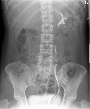
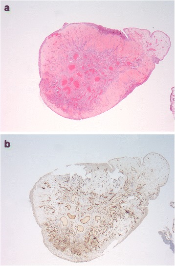
Case presentation: We present an additional, fourth case of an AVM of the ureter, clinically presented as asymptomatic haematuria and an obstructive process in the left ureter. Ureteroscopic evaluation revealed a fibroepithelial polypoid-like lesion in the proximal ureter. After biopsy showed a benign lesion, the lesion was treated with the 2-μm continuous wave (cw) thulium laser. Histopathological examination revealed a polypoid laesion caused by a circumscribed arteriovenous malformation. Almost four years after operation the patient remains asymptomatic and free of recurrence.
Conclusion: Arteriovenous malformations of the urinary tract are extremely rare. We presented a fourth case of a arteriovenous malformation of the ureter.
Keywords: Arteriovenous malformation; Case report; Polyp; Ureter.
Conflict of interest statement
Ethics approval and consent to participate
Not applicable.
Consent for publication
We received written consent for publication from the patient.
Competing interests
The authors declare that they have no competing interests.
Publisher’s Note
Springer Nature remains neutral with regard to jurisdictional claims in published maps and institutional affiliations.
Figures
References
-
- Sun Y, Xu C, Wen X, et al. Is endoscopic management suitable for long ureteral fibroepithelial polyps? J Endourol. 2008;22:1459–62. http://online.liebertpub.com.proxy.library.uu.nl/doi/abs/10.1089/end.200.... - DOI - PubMed
-
- Sech SM, Saboorian MH, Ashfaq R, et al. Polypoid arteriovenous malformation of the ureter. J Urol. 1997;158:1903–4. http://www.jurology.com/article/S0022-5347(01)64166-1/fulltext. - PubMed
Publication types
MeSH terms
LinkOut - more resources
Full Text Sources
Other Literature Sources
Medical
Molecular Biology Databases
