

Interaction of γ-Fe2O3 nanoparticles with Citrus maxima leaves and the corresponding physiological effects via foliar application
- PMID: 28693496
- PMCID: PMC5504858
- DOI: 10.1186/s12951-017-0286-1
Interaction of γ-Fe2O3 nanoparticles with Citrus maxima leaves and the corresponding physiological effects via foliar application
Abstract
Background: Nutrient-containing nanomaterials have been developed as fertilizers to foster plant growth and agricultural yield through root applications. However, if applied through leaves, how these nanomaterials, e.g. γ-Fe2O3 nanoparticles (NPs), influence the plant growth and health are largely unknown. This study is aimed to assess the effects of foliar-applied γ-Fe2O3 NPs and their ionic counterparts on plant physiology of Citrus maxima and the associated mechanisms.
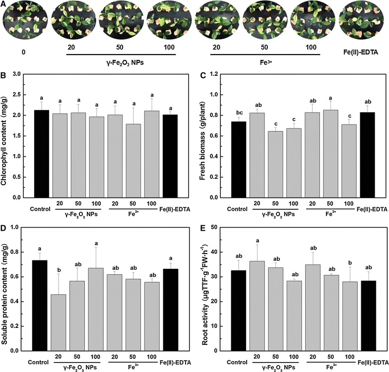
Results: No significant changes of chlorophyll content and root activity were observed upon the exposure of 20-100 mg/L γ-Fe2O3 NPs and Fe3+. In C. maxima roots, no oxidative stress occurred under all Fe treatments. In the shoots, 20 and 50 mg/L γ-Fe2O3 NPs did not induce oxidative stress while 100 mg/L γ-Fe2O3 NPs did. Furthermore, there was a positive correlation between the dosages of γ-Fe2O3 NPs and Fe3+ and iron accumulation in shoots. However, the accumulated iron in shoots was not translocated down to roots. We observed down-regulation of ferric-chelate reductase (FRO2) gene expression exposed to γ-Fe2O3 NPs and Fe3+ treatments. The gene expression of a Fe2+ transporter, Nramp3, was down regulated as well under γ-Fe2O3 NPs exposure. Although 100 mg/L γ-Fe2O3 NPs and 20-100 mg/L Fe3+ led to higher wax content, genes associated with wax formation (WIN1) and transport (ABCG12) were downregulated or unchanged compared to the control.
Conclusions: Our results showed that both γ-Fe2O3 NPs and Fe3+ exposure via foliar spray had an inconsequential effect on plant growth, but γ-Fe2O3 NPs can reduce nutrient loss due to their the strong adsorption ability. C. maxima plants exposed to γ-Fe2O3 NPs and Fe3+ were in iron-replete status. Moreover, the biosynthesis and transport of wax is a collaborative and multigene controlled process. This study compared the various effects of γ-Fe2O3 NPs, Fe3+ and Fe chelate and exhibited the advantages of NPs as a foliar fertilizer, laying the foundation for the future applications of nutrient-containing nanomaterials in agriculture and horticulture. Graphical abstract γ-Fe2O3 NPs exposed on plants via foliar spray and genes associated with the absorption and transformation of iron, as well as wax synthesis and secretion in Citrus maxima leaves.
Keywords: Foliar spray; Gene expression; Nano-enabled fertilizer; Wax; γ-Fe2O3 nanoparticles.
Figures


References
-
- Rengel Z, Batten GD, Crowley DE. Agronomic approaches for improving the micronutrient density in edible portions of field crops. Field Crop Res. 1999;60:27–40. doi: 10.1016/S0378-4290(98)00131-2. - DOI
-
- Wallace GA, Wallace A. Micronutrient uptake by leaves from foliar sprays of EDTA chelated metals. In: Nelson SD, editor. Iron nutrition and interactions in plants. Basel: Marcel Dekker; 1982. pp. 975–978.
-
- Peñaloza JP, Márquez-Miranda V, Cabaña-Brunod M, Reyes-Ramírez R, Llancalahuen FM, Vilos C, et al. Intracellular trafficking and cellular uptake mechanism of PHBV nanoparticles for targeted delivery in epithelial cell lines. J Nanobiotechnol. 2017;15(1):1–15. doi: 10.1186/s12951-016-0241-6. - DOI - PMC - PubMed
MeSH terms
Substances
LinkOut - more resources
Full Text Sources
Other Literature Sources
