

Pancreatic metastasis from invasive pleomorphic lobular carcinoma of the breast: a rare case report
- PMID: 28693516
- PMCID: PMC5504649
- DOI: 10.1186/s13000-017-0641-4
Pancreatic metastasis from invasive pleomorphic lobular carcinoma of the breast: a rare case report
Abstract
Background: Invasive pleomorphic lobular carcinoma (PLC) is an aggressive subtype of invasive lobular carcinoma of the breast, which has its own histopathological and biological features. The metastatic patterns for PLC are distinct from those of invasive ductal carcinoma. In addition, pancreatic metastasis from PLC is extremely rare.
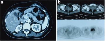
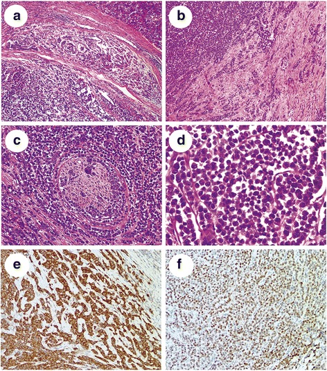
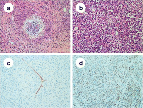
Case presentation: We report a rare case of a 48-year-old woman presenting with clinical gastrointestinal symptoms and pancreatic metastasis of PLC. The pancreatic tumor was composed of pleomorphic tumor cells arranged in the form of solid sheets and nests and as single files, with frequent mitotic figures, nucleolar prominence, high nuclear to cytoplasmic ratio and loss of cohesion. The malignant cells were positive for p120 (cytoplasmic) and GATA3 and negative for estrogen receptor, progesterone receptor, human epidermal growth factor receptor 2, E-cadherin, gross cystic disease fluid protein 15 and mammaglobin, which indicated a lobular carcinoma phenotype of the breast.
Conclusions: To the best of our knowledge, this is one of the few reported cases in the literature of pancreatic metastasis of invasive lobular carcinoma of the breast, of which the definitive diagnosis was obtained only after surgery. Rare metastasis sites should be considered, particularly, when a patient has a medical history of PLC.
Keywords: GATA3; Metastasis; Pleomorphic lobular carcinoma; Triple-negative breast cancer.
Conflict of interest statement
Ethics approval and consent to participate
This case study was approved by the Institutional Review Board for ethical committee of Fudan University Shanghai Cancer Center.
Consent for publication
Written informed consent was obtained from the patient for publication of this case report and any accompanying images. A copy of the written consent is available for review by the Editor-in-Chief of this journal.
Competing interests
The authors declare no conflicts of interest.
Publisher’s Note
Springer Nature remains neutral with regard to jurisdictional claims in published maps and institutional affiliations.
Figures
References
Publication types
MeSH terms
Substances
LinkOut - more resources
Full Text Sources
Other Literature Sources
Medical
Research Materials
