. 2017 Aug 15;144(16):2914-2924.
doi: 10.1242/dev.150227. Epub 2017 Jul 10.

PLCζ is the physiological trigger of the Ca2+ oscillations that induce embryogenesis in mammals but conception can occur in its absence

Affiliations

PLCζ is the physiological trigger of the Ca2+ oscillations that induce embryogenesis in mammals but conception can occur in its absence

Alaa Hachem et al. Development. .

Abstract

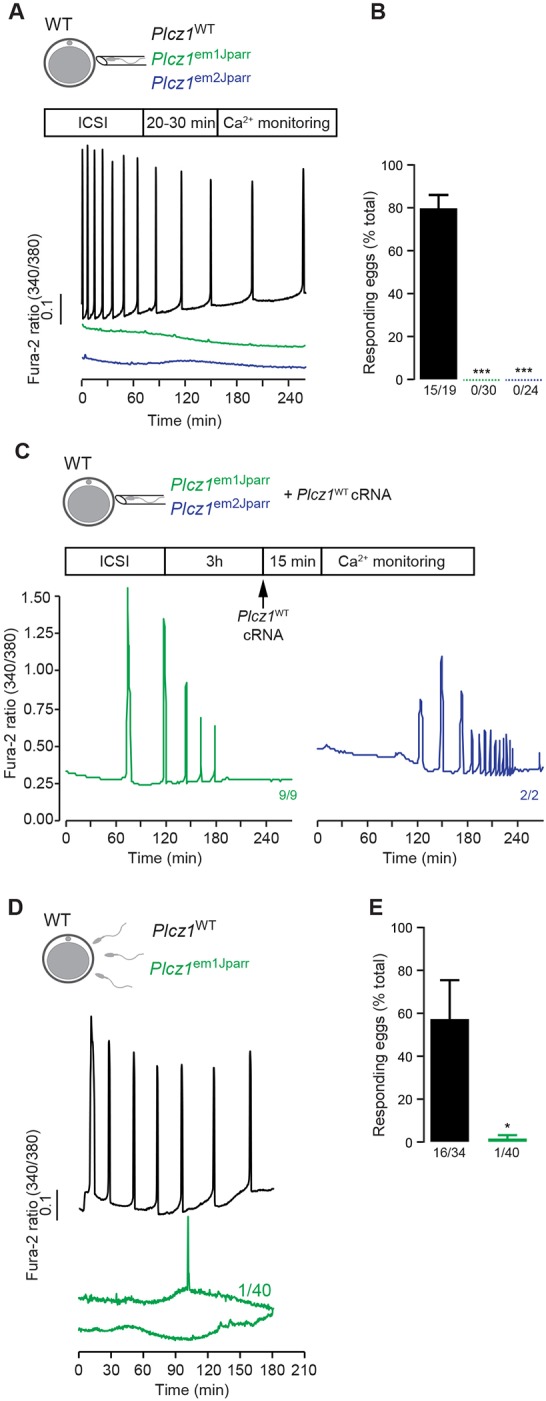
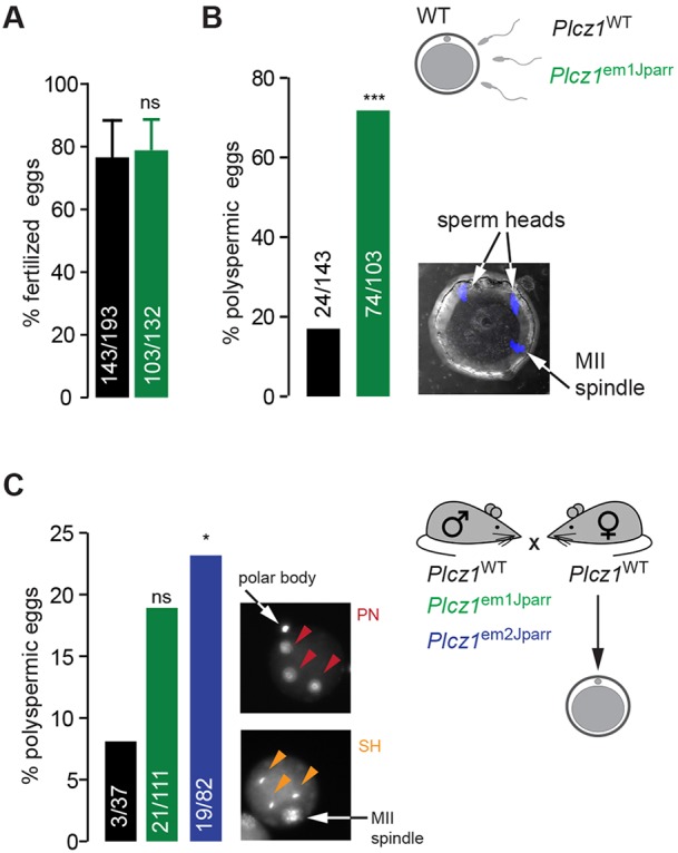
Activation of the egg by the sperm is the first, vital stage of embryogenesis. The sperm protein PLCζ has been proposed as the physiological agent that triggers the Ca2+ oscillations that normally initiate embryogenesis. Consistent with this, recombinant PLCζ induces Ca2+ oscillations in eggs and debilitating mutations in the PLCZ1 gene are associated with infertility in men. However, there has been no evidence that knockout of the gene encoding PLCζ abolishes the ability of sperm to induce Ca2+ oscillations in eggs. Here, we show that sperm derived from Plcz1-/- male mice fail to trigger Ca2+ oscillations in eggs, cause polyspermy and thus demonstrate that PLCζ is the physiological trigger of these Ca2+ oscillations. Remarkably, some eggs fertilized by PLCζ-null sperm can develop, albeit at greatly reduced efficiency, and after a significant time-delay. In addition, Plcz1-/- males are subfertile but not sterile, suggesting that in the absence of PLCζ, spontaneous egg activation can eventually occur via an alternative route. This is the first demonstration that in vivo fertilization without the normal physiological trigger of egg activation can result in offspring. PLCζ-null sperm now make it possible to resolve long-standing questions in fertilization biology, and to test the efficacy and safety of procedures used to treat human infertility.

Keywords: Calcium signalling; Egg; Embryogenesis; Mouse; PLCζ; Sperm.


Conflict of interest statement

Competing interestsThe authors declare no competing or financial interests.

Figures

Fig. 1.
Fig. 1.
Genomic characteristics of mouse Plcz1em1Jparr and Plcz1em2Jparr mutant alleles. (A) Gene structure of the mouse Plcz1 gene (NM_054066.4) and target sequences (red lines) for CRISPR/Cas9. Exons are represented by vertical bars (unfilled, untranslated regions; filled, coding regions). (B,C) Comparison of genomic sequences from wild-type Plcz1 (Plcz1WT) allele and two mutant Plcz1 alleles harbouring nucleotide deletions (deleted sequence shaded grey in Plcz1WT sequence). Capital letters correspond to exonic sequences and lower case letters to intronic sequences. (D) Typical result from a genotyping PCR reaction for both Plcz1 mutant lines: HET, Plcz1+/em1Jparr or Plcz1+/em2Jparr; MUT, homozygote Plcz1em1Jparr or Plcz1em2Jparr; WT, homozygote Plcz1WT.
Fig. 2.
Fig. 2.
Expression analysis of mouse Plcz1em1Jparr and Plcz1em2Jparr mutant alleles. (A,C) Comparison of cDNA sequences obtained from RT-PCR reactions using testis RNA from Plcz1WT and homozygote Plcz1em1Jparr (A) or Plcz1em2Jparr (C) animals. Codons are underlined, with amino acids indicated by their three-letter code. Numbers below amino acids correspond to the last amino acid position within the primary sequence not affected by the frameshift. The amino acid sequence after the frameshift is in grey. (B,D) Predicted protein-domain structures for wild-type mouse PLCζ protein (PLCζWT) and truncated proteins resulting from expression of mutant PLCζ em1Jparr (B) and PLCζ em2Jparr (D) alleles. (E) Immunoblotting analysis of PLCζ in sperm from Plcz1WT (two animals) and PLCζ em1Jparr (four animals) using a custom-made anti-PLCζ antibody (Kurokawa et al., 2005). The immunoreactive band corresponding to PLCζ is indicated by a red arrow. (F) Agarose gel image showing integrity of in vitro-produced cRNA. (G) Typical Ca2+ responses, monitored by the fluorescence ratio of Fura-2 in wild-type mouse eggs, elicited by injection of recombinant copy RNA (cRNA) in vitro transcribed from Plcz1WT, Plcz1em1Jparr or Plcz1em2Jparr cDNAs. Proportion of eggs showing Ca2+ oscillations: Plcz1WT ([cRNA] 1 ng/μl=10/10; [cRNA] 50 ng/μl=8/8), Plcz1em1Jparr ([cRNA] 50 ng/μl=5/5; [cRNA] 100 ng/μl=5/5) and Plcz1em1Jparr ([cRNA] 50 ng/μl=5/5; [cRNA] 100 ng/μl=5/5).
Fig. 3.
Fig. 3.
Characterization of sperm from homozygote Plcz1em1Jparr and Plcz1em2Jparr mice. (A) Histological analysis of testes sections stained with Hematoxylin/Eosin. (B) Density plots of sperm stained with SYBR14 (live sperm) and propidium iodide (PI; dead sperm) and analysed by flow cytometry. A total of 10,000 events were collected for each group. (C) Quantification of data from B. Representative stained sperm were obtained using an Olympus BX40 microscope. (D-G) Analysis of sperm motility parameters using computer-assisted sperm analysis (CASA). (H) An acrosome reaction was induced either by treatment with progesterone or ionomycin, and reacted sperm were assessed by lack of reactivity towards fluorescently labelled lectin PNA. Representative images of reacted and unreacted sperm are shown. All data are presented as average±s.e.m. of four different animals. There are no statistically significant differences.
Fig. 4.
Fig. 4.
Ca2+ responses elicited during fertilization of mouse eggs. (A) Representative traces of Ca2+ responses, monitored by the fluorescence ratio (excitation 340/380 nm) of Fura-2, in wild-type mouse eggs, elicited by ICSI with sperm from homozygote Plcz1WT, Plcz1em1Jparr or Plcz1em2Jparr males. (B) Compilation of results from all eggs subjected to ICSI. Data are presented as average±s.e.m. followed by a one-way ANOVA analysis; numbers shown underneath each bar indicate proportion of responding eggs; the following number of animals were used for each group in a total of four or five experiments: Plcz1WT (3), Plcz1em1Jparr (2) and Plcz1em2Jparr (4). (C) Representative traces of Ca2+ responses, monitored by the fluorescence ratio (excitation 340/380 nm) of Fura-2, in wild-type mouse eggs, elicited by injection of Plcz1WT cRNA after ICSI with sperm from homozygote Plcz1em1Jparr and Plcz1em2Jparr males. Numbers shown next to traces indicate proportion of responding eggs subjected to ICSI and cRNA injection. (D) Representative traces of Ca2+ responses, in wild-type mouse eggs, elicited by IVF with sperm from Plcz1WT and Plcz1em1Jparr males; the Plcz1WT control sperm were from a transgenic line of the same strain background as the Plcz1em1Jparr sperm, in which the catalytic subunit of PKA was exclusively modified in the sperm (Morgan et al., 2008). (E) Compilation of results from all eggs subjected to IVF. Data are presented as average±s.e.m. followed by Student's t-test; numbers shown underneath each bar indicate proportion of responding eggs; the following number of animals were used for each group in a total of four or five experiments: Plcz1WT (5) and Plcz1em1Jparr (1).
Fig. 5.
Fig. 5.
Increased polyspermy in eggs fertilized by Plcz1em1Jparr and Plcz1em2Jparr mutant sperm. (A,B) Wild-type eggs were stained with Hoechst 6 h post-IVF with Plcz1WT- and Plcz1em1Jparr-derived sperm (Plcz1WT: 193 eggs, 1 male; Plcz1em1Jparr: 132 eggs, 1 male). (A) Percentage of eggs showing sperm penetration as indicative of fertilization. (B) Percentage of eggs showing abnormal number (>1) of sperm heads. Data are presented as percentage of total eggs examined from three combined experiments. An example of a fertilized egg showing polyspermy is shown. (C) Number of polyspermic eggs derived from super-ovulated wild-type females fertilized in vivo by homozygote Plcz1WT, Plcz1em1Jparr or Plcz1em2Jparr males. Eggs were stained with Hoechst 18 h post-hCG treatment of females, for visualization of abnormal number of pronuclei (>2 PN) or sperm heads (>1 SH). Data are presented as percentage of total eggs examined from combined experiments (Plcz1WT, 37 eggs, 2 male; Plcz1em1Jparr, 111 eggs, 3 males; Plcz1em2Jparr, 82 eggs, 2 males) and statistical analysis performed using a 2×2 contingency table followed by a Fisher's exact test. Examples of fertilized eggs showing polyspermy are shown.
Fig. 6.
Fig. 6.
Embryo development in the absence of Ca2+ oscillations. (A) Proportion of two-cell and blastocyst embryos 2 days or 5 days post-ICSI, respectively (Plcz1WT, 40 eggs, 3 males; Plcz1em1Jparr, 36 eggs, 1 male; Plcz1em2Jparr, 47 eggs, 3 males). Plcz1WT cRNA was injected after ICSI, where indicated (Plcz1em1Jparr, 20 eggs, 1 male; Plcz1em2Jparr, 9 eggs, 2 males). Data are presented as average±s.e.m. of three to six experiments and example embryos are shown. (B,C) Eggs from super-ovulated females treated with hCG were fertilized in vivo, collected between 18 and 22 h post-hCG, and allowed to develop in vitro. Proportion of eggs showing sperm head(s) (SH), 1 pronucleus (1PN), 2 pronuclei (2PN) and 2-cell embryos at the indicated times. Examples of embryonic development stages are shown. (B) Data for 18 h were obtained from Hoechst-stained eggs (Plcz1WT, 37 eggs, 2 male; Plcz1em1Jparr, 111 eggs, 3 males; Plcz1em2Jparr, 82 eggs, 2 males). (C) Data from 22 h onwards were obtained from: Plcz1WT, 25 eggs, 2 males; Plcz1em1Jparr, 120 eggs, 3 males; Plcz1em2Jparr, 43 eggs, 1 male. For all panels, statistical significance was analysed using one-way ANOVA.

References

    1. Aarabi M., Yu Y., Xu W., Tse M. Y., Pang S. C., Yi Y.-J., Sutovsky P. and Oko R. (2012). The testicular and epididymal expression profile of PLCzeta in mouse and human does not support its role as a sperm-borne oocyte activating factor. PLoS ONE 7, e33496 10.1371/journal.pone.0033496 - DOI - PMC - PubMed
    1. Aarabi M., Balakier H., Bashar S., Moskovtsev S. I., Sutovsky P., Librach C. L. and Oko R. (2014). Sperm-derived WW domain-binding protein, PAWP, elicits calcium oscillations and oocyte activation in humans and mice. FASEB J. 28, 4434-4440. 10.1096/fj.14-256495 - DOI - PubMed
    1. Bassett A. and Liu J.-L. (2014). CRISPR/Cas9 mediated genome engineering in Drosophila. Methods 69, 128-136. 10.1016/j.ymeth.2014.02.019 - DOI - PubMed
    1. Bernhardt M. L., Lowther K. M., Padilla-Banks E., McDonough C. E., Lee K. N., Evsikov A. V., Uliasz T. F., Chidiac P., Williams C. J. and Mehlmann L. M. (2015). Regulator of G-protein signaling 2 (RGS2) suppresses premature calcium release in mouse eggs. Development 142, 2633-2640. 10.1242/dev.121707 - DOI - PMC - PubMed
    1. Bianchi E., Doe B., Goulding D. and Wright G. J. (2014). Juno is the egg Izumo receptor and is essential for mammalian fertilization. Nature 508, 483-487. 10.1038/nature13203 - DOI - PMC - PubMed

Publication types