. 2017 Jul 10;7(1):5017.
doi: 10.1038/s41598-017-01772-7.

Interactome and reciprocal activation of pathways in topical mesenchymal stem cells and the recipient cerebral cortex following traumatic brain injury

Affiliations

Interactome and reciprocal activation of pathways in topical mesenchymal stem cells and the recipient cerebral cortex following traumatic brain injury

Ping K Lam et al. Sci Rep. .

Abstract

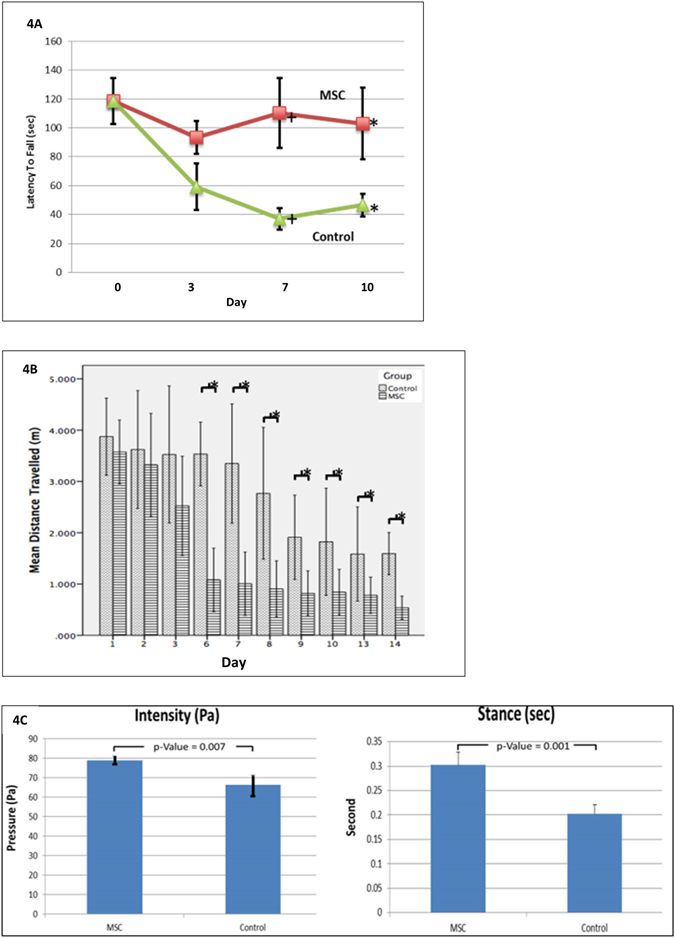
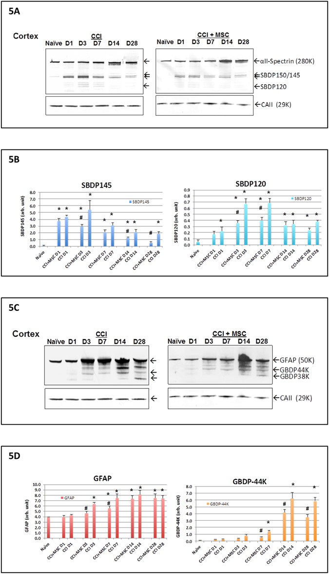
In this study, GFP-MSCs were topically applied to the surface of cerebral cortex within 1 hour of experimental TBI. No treatment was given to the control group. Three days after topical application, the MSCs homed to the injured parenchyma and improved the neurological function. Topical MSCs triggered earlier astrocytosis and reactive microglia. TBI penumbra and hippocampus had higher cellular proliferation. Apoptosis was suppressed at hippocampus at 1 week and reduced neuronal damaged was found in the penumbral at day 14 apoptosis. Proteolytic neuronal injury biomarkers (alphaII-spectrin breakdown products, SBDPs) and glial cell injury biomarker, glial fibrillary acidic protein (GFAP)-breakdown product (GBDPs) in injured cortex were also attenuated by MSCs. In the penumbra, six genes related to axongenesis (Erbb2); growth factors (Artn, Ptn); cytokine (IL3); cell cycle (Hdac4); and notch signaling (Hes1) were up-regulated three days after MSC transplant. Transcriptome analysis demonstrated that 7,943 genes were differentially expressed and 94 signaling pathways were activated in the topical MSCs transplanted onto the cortex of brain injured rats with TBI. In conclusion, topical application offers a direct and efficient delivery of MSCs to the brain.


Conflict of interest statement

The authors declare that they have no competing interests.

Figures

Figure 1
Figure 1
MSC phenotype and characterization in culture. An image of GFP-MSCs in culture showed a spindle-shaped morphology (1A), phase-contrast microscopy, x100). Flow cytometric analysis of MSCs using phycoerythrin-conjugated anti-CD29, anti-CD90 and anti-CD45 (1B).
Figure 2
Figure 2
Fate of GFP+ve MSCs transplanted onto penumbra cortex following TBI. Few GFP+ve cells (arrows) were found in the penumbral region of TBI 3 days after topical application. (2A Immunohistochemistry staining IHC x200; 2B H&E x200). A TBI lesion without treatment (2C H&E x200). The homed MSCs which were pre-labeled with CM-DIL red fluorescence dye expressed markers of GFAP (Green) (2D Immunofluorescent staining IF x200); Nestin (Green) (2E IF x200) and NeuN (Green) (2F IF x200). CXCR4 (Red) was expressed by the homed MSCs (Green) (2 G IF x200). In the vicinity of homed MSCs (Green), the TBI penumbra expressed SDF-1 (Red) (2 H IF x200). DAPI (Blue) was used to stain the nucleus.
Figure 3
Figure 3
Representative photos of histochemical staining images at different time points post-MSC treatment. At day 3, MSC-treatment group had more GFAP+ve cells in the TBI penumbra (3A, IHC x400) and hippocampus (3B IHC x400). More Iba-1+ve cells (3C HIC x400) were observed in the penumbra of MSC-treatment group. Both the penumbra (3D IHC x400) and hippocampus (3D IHC x400) also had higher PCNA expression. MSCs attenuated apoptosis in the hippocampus (3F Tunel, x400). At day 14, more neurons were found in both penumbra and hippocampus of MSC-treatment group (3G Cresyl Violet x400).
Figure 4
Figure 4
Behavioral assessment of rats with and without MSC treatment following TBI. Rats treated with topical MSCs showed a significant improvement in behavioral assessment. Parameters were expressed in mean + standard deviation. Topical MSCs improved the coordination and integration of movement motor function in the Motor Rod Test (4A). The animals travelled a shorter distance to reach the platform in the Water Maze Test (4B). Automated gait analysis illustrated the greater intensity (mean pressure exerted on the floor by one individual paw) and longer stance (duration during which the paw is in contact with the glass plate) by the MSC-treatment group (4C).
Figure 5
Figure 5
Expression of biomarkers in penumbra cortex in rats with and without MSC treatment after TBI. Effects of MSC topical transplantation on proteolytic neuronal and glial injury biomarker levels in the ipsilateral cortex axonal alphaII-spectrin breakdown product SBDP biomarkers levels at different time points after controlled cortical impact (CCI) injury with or without MSC topical transplant are shown. (A) showed representative blots of spectrin and house-keeping gene loading control (Carbonic anhydrase II, 29 kDa). (B) showed quantification of biomarkers (equalized by CA-II band intensity). GFAP and GFAP-breakdown product GBDP biomarkers levels at different time points after controlled cortical impact (CCI) injury with or without MSC topical transplant were shown (C,D) shows representative blots of GFAP and house-keeping gene (Carbonic anhydrase II, 29 kDa). Quantification of comparison: *higher than naïve group; #MSC + CCI treatment group was different from CCI alone group at same time point (p < 0.05). MSC + CCI vs. CCI group-based ANOVA results were stated in the text.
Figure 6
Figure 6
Analysis of gene of MSC and penumbra cortex at day 3 following TBI. Hierarchical clustering of gene expression of MSC pellets before and after topical application on TBI brain and normal brain. Gene lists were generated by comparing MSC pellet versus topical MSCs on TBI brain and topical MSCs on normal brain using moderate T-test and 2-fold filtering in GeneSpring. Down-regulated genes were shown in blue and up-regulated genes were shown in red (6A). Venn diagram illustrated the number of genes which were up- or down-regulated of topical MSCs on TBI brain and normal brain using moderate T-test and 2-fold filtering in GeneSpring. Overlapping area indicated genes from MSCs pellets were coherently altered in both conditions (6B). Cluster analysis of PCR-array on 84 genes associated with neurogenesis in penumbral cortex treated topical MSCs (6C). Compared with control, 13 genes were significantly up-regulated (p < 0.05) (6D). 6 out of 13 genes were up-regulated with equal or more than 1.5 fold (6E). Diagram of the six-gene network which was proposed to get involved in the neurogenesis mediated by the topical MSCs. Grey color genes participated in the network but they were not detected by the PCR-array (6F).

References

    1. Mass AIR, Stocchetti N, Bullock R. Moderate and severe traumatic brain injury in adults. Lancet Neurol. 2008;7:728–741. doi: 10.1016/S1474-4422(08)70164-9. - DOI - PubMed
    1. Raghupathi R. Cell death mechanisms following traumatic brain injury. Brain Pathol. 2004;14:215–222. doi: 10.1111/j.1750-3639.2004.tb00056.x. - DOI - PMC - PubMed
    1. Das M, Mohapatra S, Mophapatra SS. New perspectives on central and peripheral immune responses to acute traumatic brain injury. J Neuroinflammation. 2012;9:236–249. doi: 10.1186/1742-2094-9-236. - DOI - PMC - PubMed
    1. Prockop DJ, et al. Defining the risks of mesenchymal stromal cell therapy. Cytotherapy. 2010;12:576–578. doi: 10.3109/14653249.2010.507330. - DOI - PubMed
    1. Rubio D, et al. Spontaneous human adult stem cell transformation. Cancer Res. 2005;65:3035–3039. - PubMed

Publication types