. 2017 Jul 10;7(1):4944.
doi: 10.1038/s41598-017-05095-5.

Analysis of positive and negative allosteric modulation in metabotropic glutamate receptors 4 and 5 with a dual ligand

Affiliations

Analysis of positive and negative allosteric modulation in metabotropic glutamate receptors 4 and 5 with a dual ligand

James A R Dalton et al. Sci Rep. .

Abstract

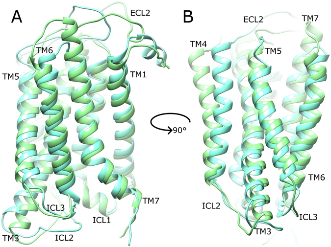
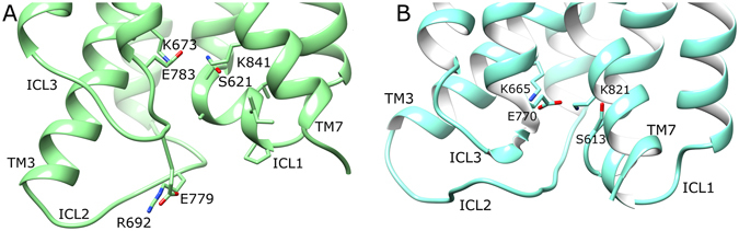
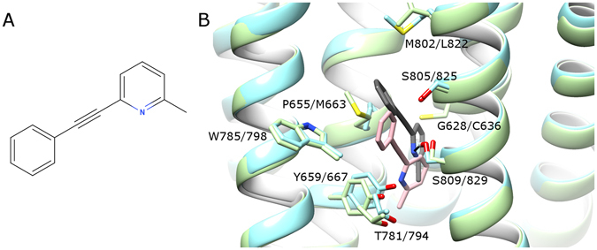
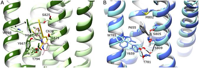
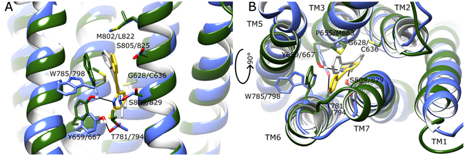
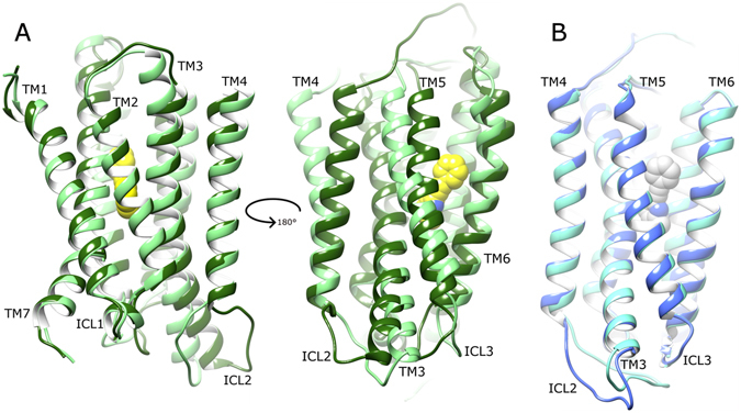
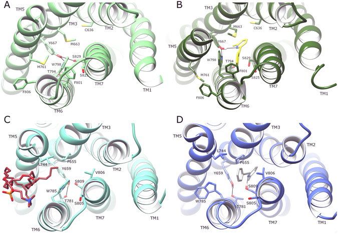
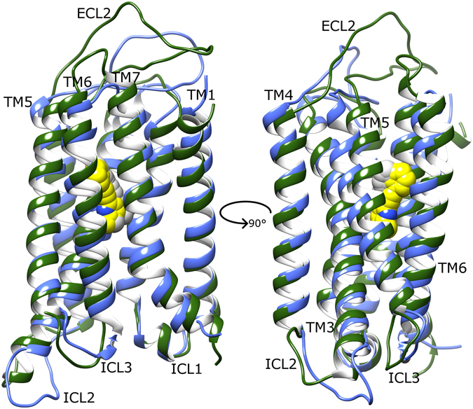
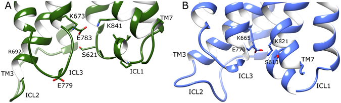
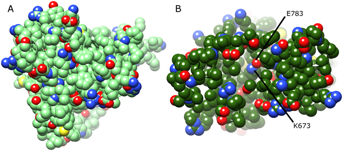
As class C GPCRs and regulators of synaptic activity, human metabotropic glutamate receptors (mGluRs) 4 and 5 are prime targets for allosteric modulation, with mGlu5 inhibition or mGlu4 stimulation potentially treating conditions like chronic pain and Parkinson's disease. As an allosteric modulator that can bind both receptors, 2-Methyl-6-(phenylethynyl)pyridine (MPEP) is able to negatively modulate mGlu5 or positively modulate mGlu4. At a structural level, how it elicits these responses and how mGluRs undergo activation is unclear. Here, we employ homology modelling and 30 µs of atomistic molecular dynamics (MD) simulations to probe allosteric conformational change in mGlu4 and mGlu5, with and without docked MPEP. Our results identify several structural differences between mGlu4 and mGlu5, as well as key differences responsible for MPEP-mediated positive and negative allosteric modulation, respectively. A novel mechanism of mGlu4 activation is revealed, which may apply to all mGluRs in general. This involves conformational changes in TM3, TM4 and TM5, separation of intracellular loop 2 (ICL2) from ICL1/ICL3, and destabilization of the ionic-lock. On the other hand, mGlu5 experiences little disturbance when MPEP binds, maintaining its inactive state with reduced conformational fluctuation. In addition, when MPEP is absent, a lipid molecule can enter the mGlu5 allosteric pocket.


Conflict of interest statement

The authors declare that they have no competing interests.

Figures

Figure 1
Figure 1
(a) Two agonists A1 and A2 activate the receptor R producing effects 1 and 2, respectively. (b) Agonist A activates receptors 1 and 2 producing effects 1 and 2, respectively. In the present study ligand A corresponds to MPEP whereas receptors 1 and 2 correspond to mGlu4 and mGlu5, respectively.
Figure 2
Figure 2
Average conformation of mGlu4 (light green) and mGlu5 (cyan) in their apo states, obtained from respective MD simulations, showing a 90° rotation between (A) and (B) around the membrane plane (extracellular-side: top, intracellular-side: bottom). Average receptor states are calculated from respective MD simulations (between 2.5–5 µs). Relevant structural features are labelled: extracellular loops (ECLs), intracellular loops (ICLs) and transmembrane helices (TMs).
Figure 3
Figure 3
Average conformations of intracellular loops and selected charged/polar residues of (A) apo mGlu4 (green) and (B) apo mGlu5 (cyan), obtained from 2.5–5 µs of respective MD simulations. Relevant structural features are labelled, i.e. intracellular loops (ICLs) and intramolecular locks, i.e. K673-E783 in mGlu4 ionic-lock and K665-E770 in mGlu5 ionic-lock.
Figure 4
Figure 4
(A) 2-Methyl-6-(phenylethynyl)pyridine (MPEP), (B) comparison of MPEP (grey) docking in mGlu5 (light blue), and MPEP (pink) docking in mGlu4 (light green) prior to MD simulations. Selected residues delineating allosteric pockets or participating in ligand binding are labelled (labelling: mGlu5 first, mGlu4 second). Protein-ligand H-bonds are represented by black lines. Helix orientations as follows: TM5 left, TM3 centre-left, TM7 centre-right, TM1 right. TM6 backbone is hidden.
Figure 5
Figure 5
Comparison of bound MPEP in (A) mGlu4 and (B) mGlu5, before and after respective MD simulations (average conformations calculated from 2.5–5 µs). Colours as follows: (A) MPEP before MD in pink, MPEP after MD in yellow, mGlu4 before MD in light green, mGlu4 after MD in dark green; (B) MPEP before MD in dark grey, MPEP after MD in light grey, mGlu5 before MD in light blue, mGlu5 after MD in blue. Selected residues delineating allosteric pockets or participating in ligand binding are labelled. Protein-ligand H-bonds are represented by solid black lines. The inward movement of W798 in mGlu4 is indicated with a dotted arrow. Helix orientations as follows: TM5 left, TM3 centre-left, TM7 centre-right, TM1 right. TM6 backbone is hidden.
Figure 6
Figure 6
A comparison of average conformations of mGlu4 with bound MPEP (green, yellow, respectively) and mGlu5 with bound MPEP (blue, grey, respectively) from 2.5–5 µs of respective MD simulations from following perspectives: (A) side-view within membrane and (B) extracellular top-view. Selected residues delineating allosteric pockets or participating in ligand binding are labelled (mGlu5 first, mGlu4 second). Protein-ligand H-bonds are represented by black lines.
Figure 7
Figure 7
Comparison of average conformations of (A) mGlu4 with bound MPEP (dark green, yellow, respectively) and apo mGlu4 without MPEP (light green) and (B) mGlu5 with bound MPEP (blue, light grey, respectively) and apo mGlu5 without MPEP (cyan), obtained from 2.5–5 µs of respective MD simulations. A 180° rotation around the membrane plane of mGlu4 allows differences to be noted from opposite angles. Receptors are viewed from side, within membrane, with extracellular-side, top, and intracellular-side, bottom. Relevant structural features are labelled, i.e. intracellular loops (ICLs) and transmembrane helices (TMs).
Figure 8
Figure 8
Comparison of average conformations of (A) apo mGlu4 without MPEP (light green), (B) mGlu4 with bound MPEP (dark green, yellow, respectively), (C) apo mGlu5 without MPEP (cyan) with bound lipid molecule (brown), (D) mGlu5 with bound MPEP (blue, light grey, respectively), each obtained from 2.5–5 µs of respective MD simulations. Receptors are viewed extracellular-side from top. Relevant structural features are labelled, i.e. transmembrane helices (TMs) and residues in the allosteric pocket interacting with lipid/MPEP.
Figure 9
Figure 9
Comparison of average conformations of (A) mGlu4 with bound with MPEP (dark green, yellow, respectively) and mGlu5 bound with MPEP (blue, grey, respectively), obtained from 2.5–5 µs of respective MD simulations. Receptors are viewed from side, within membrane, with a 90° rotation around membrane plane for alternative views (left and right images). Relevant structural features are labelled, i.e. intracellular loops (ICLs) and transmembrane helices (TMs).
Figure 10
Figure 10
Average conformations of intracellular loops and selected charged/polar residues of (A) MPEP-bound mGlu4 (green) and (B) MPEP-bound mGlu5 (blue), obtained from 2.5–5 µs of respective MD simulations. Relevant structural features and residues are labelled, i.e. intracellular loops (ICLs), and ionic-locks: K673-E783 in mGlu4 and K665-E770 in mGlu5.
Figure 11
Figure 11
Comparison of average conformations of (A) apo mGlu4 without MPEP (green) and (B) mGlu4 with bound MPEP (dark green) obtained from 2.5–5 µs respective MD simulations. Receptors are viewed from their intracellular side with atoms displayed as spheres. Residues participating in the ionic-lock are labelled in (B).

References

    1. Pin JP, Acher F. The metabotropic glutamate receptors: structure, activation mechanism and pharmacology. Current drug targets. CNS and neurological disorders. 2002;1:297–317. doi: 10.2174/1568007023339328. - DOI - PubMed
    1. Pin JP, Galvez T, Prezeau L. Evolution, structure, and activation mechanism of family 3/C G-protein-coupled receptors. Pharmacology & therapeutics. 2003;98:325–354. doi: 10.1016/S0163-7258(03)00038-X. - DOI - PubMed
    1. Kniazeff J, Prezeau L, Rondard P, Pin JP, Goudet C. Dimers and beyond: The functional puzzles of class C GPCRs. Pharmacology & therapeutics. 2011;130:9–25. doi: 10.1016/j.pharmthera.2011.01.006. - DOI - PubMed
    1. Binet V, et al. Common structural requirements for heptahelical domain function in class A and class C G protein-coupled receptors. The Journal of biological chemistry. 2007;282:12154–12163. doi: 10.1074/jbc.M611071200. - DOI - PMC - PubMed
    1. Goudet C, et al. Heptahelical domain of metabotropic glutamate receptor 5 behaves like rhodopsin-like receptors. Proceedings of the National Academy of Sciences of the United States of America. 2004;101:378–383. doi: 10.1073/pnas.0304699101. - DOI - PMC - PubMed

Publication types

Substances