. 2017 Jul 10;7(1):5000.
doi: 10.1038/s41598-017-04977-y.

Presence of diabetes autoantigens in extracellular vesicles derived from human islets

Affiliations

Presence of diabetes autoantigens in extracellular vesicles derived from human islets

Craig P Hasilo et al. Sci Rep. .

Abstract

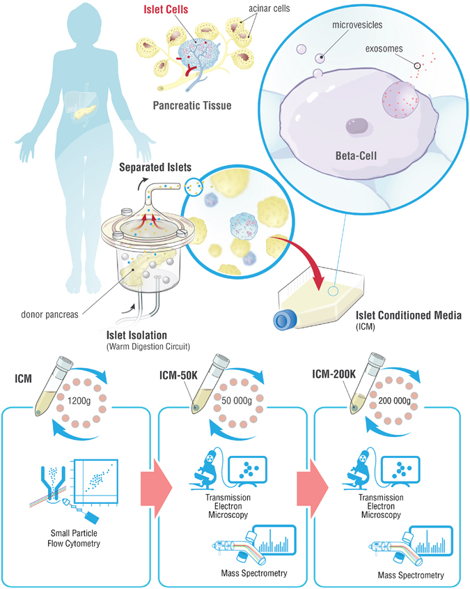
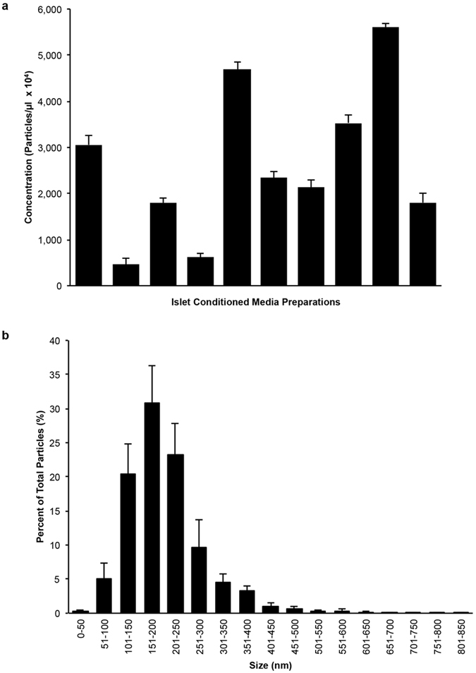
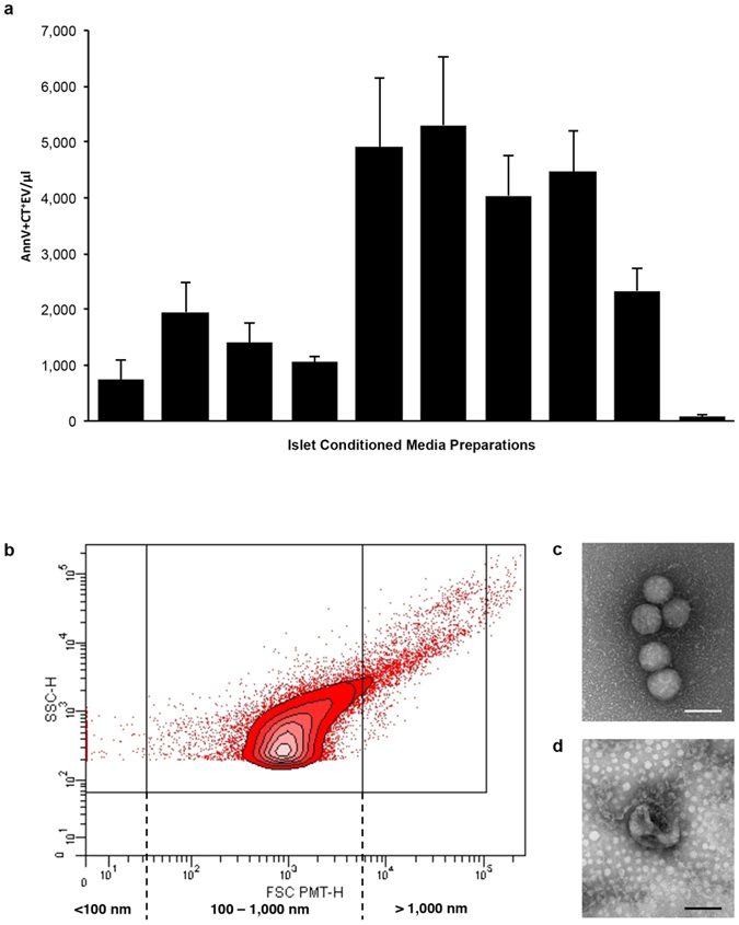
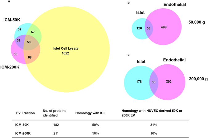
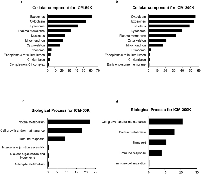
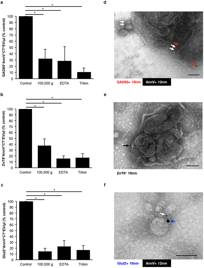
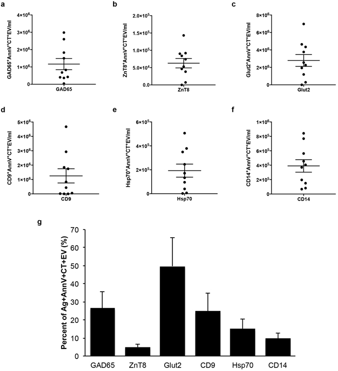
Beta-cell (β-cell) injury is the hallmark of autoimmune diabetes. However, the mechanisms by which autoreactive responses are generated in susceptible individuals are not well understood. Extracellular vesicles (EV) are produced by mammalian cells under normal and stressed physiological states. They are an important part of cellular communication, and may serve a role in antigen processing and presentation. We hypothesized that isolated human islets in culture produce EV that contain diabetes autoantigens (DAA) from these otherwise normal, non-diabetic donors. Here we report the caspase-independent production of EV by human islets in culture, and the characterization of DAA glutamic acid decarboxylase 65 (GAD65) and zinc transporter 8 (ZnT8), as well as the β-cell resident glucose transporter 2 (Glut2), present within the EV.


Conflict of interest statement

The authors declare that they have no competing interests.

Figures

Figure 1
Figure 1
Schema describing the workflow to generate and analyze human islet conditioned media extracellular vesicles. Islet isolation was performed after the surgical retrieval of a donor pancreas. The pancreas was enzymatically digested in a closed loop circuit until free islets were observed. The islets were then purified, and cultured for up to 72 hours. Islet conditioned medium (ICM) was harvested, centrifuged at 1,200 × g for 15 min. and analyzed by small particle flow cytometry for DAA and markers of EV, or serially centrifuged a 50,000 × g and 200,000 × g for analysis by transmission electron microscopy or mass spectrometry on the ICM-50 K and ICM-200 K fractions.
Figure 2
Figure 2
Cultured human islets produce distinct populations of particles. (a) Nanoparticle tracking analysis (NTA) graph of mean particle levels in ICM from ten donors (n = 10) shows variable levels between preparations. (b) NTA histogram of particle size distribution shows the majority of particles detected are between 100 nm and 650 nm. Data are expressed as mean ± SEM of ten donors (n = 10). At least 5 recordings, 30 seconds each were obtained for each sample at 37 °C, camera shutter speed of 30.0 ms, camera level 14 and detection threshold set to 9.
Figure 3
Figure 3
Human islet conditioned media contains a heterogeneous population of AnnV+CellTracker+EV. (a) Bar graph showing the mean levels of each ICM (n = 10) detected by flow cytometry varies in their content of AnnV+Cell Tracker+EV after 24 hours in culture. (b) Representative contour plot (FSC vs SSC) shows the peak density of AnnV+Cell Tracker+EV is within 100 nm to 1,000 nm size based on the detection of 100, 500 and 1,000 nm silica beads. (c,d) Visualization of fractionated ICM by whole mount negative staining TEM confirms the presence of membrane vesicles. Scale bars represent 100 nm. Data are expressed as mean ± SEM of n = 3 independent experiments performed on ICM from ten (n = 10) different donors.
Figure 4
Figure 4
Proteomic analysis reveals homology of islet conditioned media extracellular vesicles with islet cell lysate, and a signature distinct from extracellular vesicles produced by human umbilical vein endothelial cells. Venn diagrams depicting the overlap of genes detected in the islet conditioned media (ICM)-50 K and ICM-200 K fractions with that of the islet cell lysate (ICL) (a). Comparison of homology of genes detected in ICM-50 K compared to that of human umbilical vein endothelial cell (HUVEC)-50 K, as well as in ICM-200 K and HUVEC-200 K (islet and endothelial 50,000 g (b) and 200,000 g (c), respectively).
Figure 5
Figure 5
Comparison of proteomic data by enrichment analyses of fractionated human islet conditioned media with FunRich component and pathway analyses. Bar graphs of cellular compontments overrepresented in highly abundant proteins are shown for islet conditioned media (ICM)-50 K (a) and ICM-200 K (b). Bar graphs illustrating the biological process overrepresented in highly abundant proteins for ICM-50 K (c) and ICM-200 K (d).
Figure 6
Figure 6
Diabetes autoantigens and β-cell markers are present on extracellular vesicles produced by non-diabetic donors. (ac) Bar graphs of islet conditioned media (ICM) compared to signal depletion by 100,000 g centrifugation, 50 mM EDTA, or 0.3% Triton X-100 for GAD65+AnnV+CT+EV (a), ZnT8+AnnV+CT+EV (b), and Glut2+AnnV+CT+EV (c). (df) Micrographs of immunogold double labeled islet-derived EV preparations from fractionated ICM confirms the presence of AnnV (white arrows), GAD65 ((b) red arrows), ZnT8 ((d) black arrow) and Glut2 ((f) blue arrow). Data are expressed as mean ± SEM from n = 4 human islet preparations from different donors. *p < 0.05, repeated measures one-way analysis of variance with Tukey’s post-hoc test performed on raw values prior to conversion to percent of control. Scale bars represent 100 nm.
Figure 7
Figure 7
Antigen levels on extracellular vesicles present in islet conditioned media from different donor islet preparations. Column scatter graphs of islet conditioned media (ICM) analyzed by flow cytometry for GAD65+AnnV+CT+EV (a), ZnT8+AnnV+CT+EV (b), Glut2+AnnV+CT+EV (c), CD9+AnnV+CT+EV (d), Hsp70+AnnV+CT+EV (e), and CD14+AnnV+CT+EV (f). Bar graph depicting the percent of AnnV+CT+EV that were positive for the antigens described in the above panels (g). Data are expressed as mean ± SEM from n = 10 human islet preparations from different donors, and are representative of n = 3 independent experiments.

References

    1. Notkins AL, Lernmark A. Autoimmune type 1 diabetes: resolved and unresolved issues. J Clin Invest. 2001;108:1247–1252. doi: 10.1172/JCI14257. - DOI - PMC - PubMed
    1. Baekkeskov S, et al. Identification of the 64 K autoantigen in insulin-dependent diabetes as the GABA-synthesizing enzyme glutamic acid decarboxylase. Nature. 1990;347:151–156. doi: 10.1038/347151a0. - DOI - PubMed
    1. Wenzlau JM, et al. The cation efflux transporter ZnT8 (Slc30A8) is a major autoantigen in human type 1 diabetes. Proc Natl Acad Sci USA. 2007;104:17040–17045. doi: 10.1073/pnas.0705894104. - DOI - PMC - PubMed
    1. Payton MA, Hawkes CJ, Christie MR. Relationship of the 37,000- and 40,000-M(r) tryptic fragments of islet antigens in insulin-dependent diabetes to the protein tyrosine phosphatase-like molecule IA-2 (ICA512) J Clin Invest. 1995;96:1506–1511. doi: 10.1172/JCI118188. - DOI - PMC - PubMed
    1. Wenzlau JM, Hutton JC. Novel diabetes autoantibodies and prediction of type 1 diabetes. Curr Diab Rep. 2013;13:608–615. doi: 10.1007/s11892-013-0405-9. - DOI - PMC - PubMed

Publication types

MeSH terms