. 2017 Jul 10;7(1):5013.
doi: 10.1038/s41598-017-04993-y.

Revealing enterovirus infection in chronic human disorders: An integrated diagnostic approach

Affiliations

Revealing enterovirus infection in chronic human disorders: An integrated diagnostic approach

Angelo Genoni et al. Sci Rep. .

Abstract

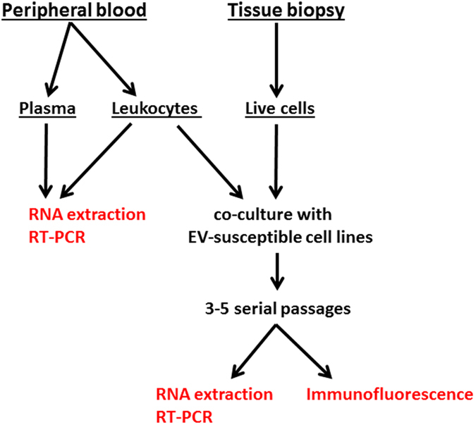
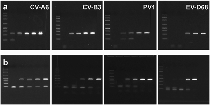
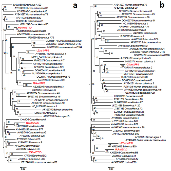
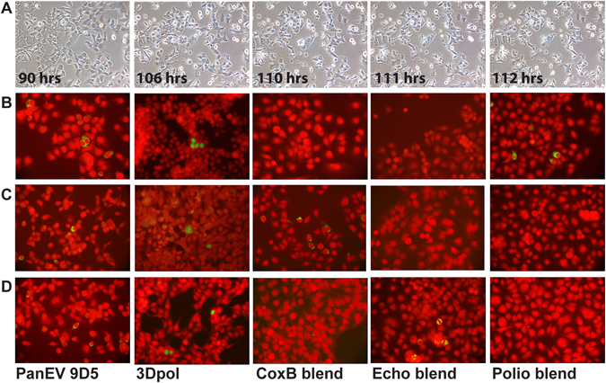
Enteroviruses (EVs) causing persisting infection are characterized by minimal replication and genetic changes. Typing of these agents may complement disease assessment and shed light on pathogenesis. Here we report an integrated approach for EV detection in human samples that is based on pre-enrichment of virus in cell culture before search for the viral genome and viral antigens. Cases of post-polio syndrome, type 1 diabetes, and chronic cardiomyopathy were investigated. As tissue-based approaches require invasive procedures, information was mainly gleaned from virus in blood. Molecular assays targeting conserved genome regions of all EV types (5'UTR, 2 C, 3Dpol) were employed. As compared to direct assays of plasma or leukocytes, the EV detection rate was significantly enhanced by co-culture of leukocytes with cell lines prior to molecular and immunologic tests. Results of RT-PCR and sequencing were confirmed by staining cell cultures with a panel of EV-specific antibodies. Sequence and phylogenetic analysis showed that EVs of the C species (polioviruses) were associated with the post-polio syndrome, while members of the B species were found in type 1 diabetes and cardiomyopathy. The procedure may be used for investigating the possible association of different EVs with a variety of chronic neurologic, endocrine, and cardiac disorders.


Conflict of interest statement

The authors declare that they have no competing interests.

Figures

Figure 1
Figure 1
Representation of the integrated procedure used for detecting enteroviruses (EVs) in patients diagnosed with post-polio syndrome (PPS), type-1 diabetes mellitus (T1D), chronic viral cardiomyopathy (CVC). Venous peripheral blood or tissue biopsies were used as clinical samples. Plasma and blood leukocytes were tested directly by molecular assays (left side). Blood leukocytes and live cells obtained from biopsies were co-cultured with cell lines (right side). Cell cultures were serially passaged 3 to 5 times. RNA was then extracted from medium of cultured cells. Molecular assay were used to detect the viral genome. For each case, the expression of enteroviral antigens in cultured cells was evaluated by immunofluorescence with a panel of anti-EV antibodies.
Figure 2
Figure 2
Results of end-point PCR of quantitated pU57 plasmids containing the 5′UTR region of CV-A6, CV-B3, PV-1, EV-D68 (representative enteroviruses of the A, B, C, and D species). Ten-fold dilutions of each plasmid were tested by end-point PCR with two different primer pairs: 5′UTR-Tok (panel a) and 5′UTR-C (panel b). For each gel, six lanes of cropped gels are shown (left to right): DNA size markers, PCR amplicons obtained with 1.6, 16, 160, 1,600, or 16,000 template molecules per reaction. Primer pairs were detecting ≤16 template molecules/reaction. Original, uncropped images are presented in the Supplementary Materials (Figures S1 and S2).
Figure 3
Figure 3
Phylogenetic analysis of nucleotide sequences of a part of the 5′-UTR region (nucleotides 345–607, panel a) and a region covering the distal part of 5′-UTR, VP4 and the proximal part of VP2 (nucleotides 580–1170, panel b) determined for PCR amplicons. Sequences determined for PPS, T1D, and CVC cases are shown in red. Homologous regions from a selected set of 50 published reference enterovirus sequences were used to determine their relatedness using alignment-free k-mer mismatch method. Phylogenetic trees were reconstructed from the distance matrix using neighbor-joining method. The quality of each internal branch of the tree was assessed as a percentage of all possible 4-node subsets that confirmed the local topology of the tree.
Figure 4
Figure 4
Secondary structure of the IRES element domain V from PPS cases RBJ/va15 (A) and LZL/va10 (B). Hairpin structures are presented in black for viruses that are most closely related to the PCR product sequences from respective cases. Mutations in the sequences from PPS cases are shown in red. In most cases these mutations disrupt the secondary structure of domain V, suggesting that the mutated agents have reduced fitness.
Figure 5
Figure 5
Slow development of morphologic changes evidenced by time-lapse microscopy in cell cultures infected with a viral isolate obtained from a T1D case and serially passaged 4 times (Row A). Incubation times are indicated in hours. Pictures were taken at 30 minutes intervals. Death of scattered cells was first observed after >100 hr incubation. Ballooning of scattered cells started appearing at 110 hrs. Then, in a couple of hours, cells were bursting with the release of multiple small vesicles (111 and 112 hrs). Original magnification 10x. Indirect immunofluorescence of human AV3 cell monolayers that had been co-cultured with leukocytes of patients diagnosed with PPS, T1D, or CVC and serially passaged 4–6 times. A panel of MAbs against EVs was used (green staining); Evans blue counterstain (red). Original magnification 20x. (Row B) cell monolayers exposed to leukocytes of a PPS case. Panenterovirus MAb 9D5 directed to VP1 and MAb 3D-05 directed to the 3D viral RNA polymerase produced cytoplasmic or nuclear staining in 1–2 percent of cells. MAb blends against coxsackieviruses B or echoviruses failed to detect virus antigens, whereas a blend of MAbs to poliovirus-1, -2, -3 produced clear cytoplasmic fluorescence. The PPS case was associated with an EV belonging to the poliovirus group (C species). (Row C) cell monolayers exposed to leukocytes of a T1D case. MAbs 9D5 and 3D-05 produced cytoplasmic staining in 1–2 percent of cells. The MAb blend to coxsackieviruses B produced granular cytoplasmic fluorescence; no staining was obtained with MAbs to echoviruses or polioviruses. The T1D case was associated with a non-polio EV, possibly belonging to the Coxsackie B group (B species). (Row D) cell monolayers exposed to leukocytes of a CVC case. MAbs 9D5 and 3D-05 produced cytoplasmic or nuclear staining in 1–2 percent of cells. The MAb blend to coxsackieviruses B did not detect virus antigens, whereas a MAb blend to echoviruses produced granular cytoplasmic fluorescence. No staining was obtained with poliovirus MAbs. This case was associated with a non-polio EV possibly belonging to the Echovirus group (B species).

References

    1. Abedi GR, et al. Enterovirus and Human Parechovirus Surveillance - United States, 2009–2013. MMWR Morb Mortal Wkly Rep. 2015;64:940–943. doi: 10.15585/mmwr.mm6434a3. - DOI - PubMed
    1. Pallansch, M. A., Oberste, M. S. & Whitton, J. L. In Fields Virology Vol. 2 (ed. Knipe, D. M. & Howley, P. M. & Fields, B. N.) 490–530 (Wolters Kluwer Health/Lippincott Williams & Wilkins, 2013).
    1. Boettler T, von Herrath M. Protection against or triggering of Type 1 diabetes? Different roles for viral infections. Expert Rev Clin Immunol. 2011;7:45–53. doi: 10.1586/eci.10.91. - DOI - PMC - PubMed
    1. Kapusinszky B, Minor P, Delwart E. Nearly constant shedding of diverse enteric viruses by two healthy infants. J Clin Microbiol. 2012;50:3427–3434. doi: 10.1128/JCM.01589-12. - DOI - PMC - PubMed
    1. Asturias EJ, et al. Humoral and intestinal immunity induced by new schedules of bivalent oral poliovirus vaccine and one or two doses of inactivated poliovirus vaccine in Latin American infants: an open-label randomised controlled trial. Lancet. 2016;388:158–169. doi: 10.1016/S0140-6736(16)00703-0. - DOI - PubMed

Publication types

MeSH terms