

Pilomatrixoma Presenting as a Rapidly Expanding Mass of the Infant Nasion
- PMID: 28694911
- PMCID: PMC5486210
Pilomatrixoma Presenting as a Rapidly Expanding Mass of the Infant Nasion
Abstract
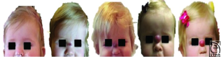
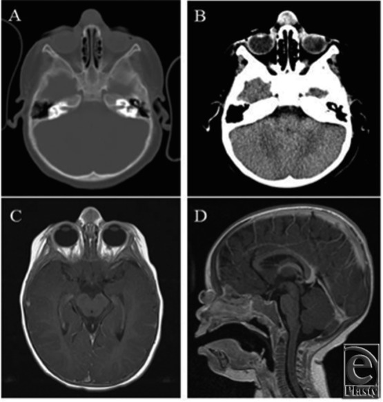
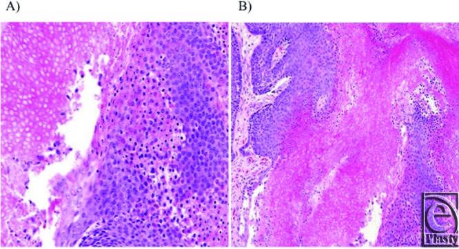
Objective: Pilomatrixomas are benign neoplasms originating from the cells of hair follicles. They typically present as a slowly enlarging, solitary mass on hair-bearing areas of the head and neck. While a common childhood lesion, pilomatrixomas are unusual in infancy. Our objective is to present an atypical pilomatrixoma located on the midline nasion of an 11-month-old as such a lesion and its management has not been previously described. Methods: Despite preoperative diagnostic imaging, including computed tomography and magnetic resonance imaging, the diagnosis was not made until examination by pathology after complete surgical excision. We also completed a thorough review of the literature pertaining to pilomatrixomas, which is presented in a concise fashion. Results: Our patient's clinical presentation did not correlate with traditional descriptions in the literature, skewing preoperative diagnosis. However, surgical management was ultimately appropriate and effective. To date, the patient has not demonstrated evidence of recurrence. Conclusion: We believe that this is the first such reported presentation of a pilomatrixoma. Given its incidence, we encourage readers to consider this diagnosis when evaluating similar pediatric skin lesions of the head and neck. Complete surgical excision is the definitive treatment.
Keywords: hair diseases; infant; midline mass; pilomatrixoma; skin neoplasms.
Figures
Similar articles
-
Pilomatrixoma and its Imitators.Ear Nose Throat J. 2024 Mar;103(3):183-189. doi: 10.1177/01455613211044778. Epub 2021 Sep 22. Ear Nose Throat J. 2024. PMID: 34549614 Review.
-
Head and neck pilomatrixoma in children.Arch Otolaryngol Head Neck Surg. 2001 Dec;127(12):1481-3. doi: 10.1001/archotol.127.12.1481. Arch Otolaryngol Head Neck Surg. 2001. PMID: 11735819
-
Clinical analysis and review of literature on pilomatrixoma in pediatric patients.Arch Craniofac Surg. 2020 Oct;21(5):288-293. doi: 10.7181/acfs.2020.00528. Epub 2020 Oct 20. Arch Craniofac Surg. 2020. PMID: 33143396 Free PMC article.
-
Pilomatrixoma: A Comprehensive Review of the Literature.Am J Dermatopathol. 2018 Sep;40(9):631-641. doi: 10.1097/DAD.0000000000001118. Am J Dermatopathol. 2018. PMID: 30119102 Review.
-
Pilomatrixoma of the head and neck in children.Otolaryngol Head Neck Surg. 2001 Nov;125(5):510-5. doi: 10.1067/mhn.2001.117371. Otolaryngol Head Neck Surg. 2001. PMID: 11700451
References
-
- Forbis R, Helwig EB. Pilomatrixoma (calcifying epithelioma) Arch Dermatol. 1961;83:606–18. - PubMed
-
- Agarwal RP, Handler SD, Matthews MR, Carpentieri D. Pilomatrixoma of the head and neck in children. Otolaryngol Head Neck Surg. 2001;125:510–15. - PubMed
-
- Noguchi H, Hayashibara T, Ono T. A statistical study of calcifying epithelioma, focusing on the sites of origin. J Dermatol. 1995;22:24–7. - PubMed
-
- Kwon D, Grekov K, Krishnan M, Dyleski R. Characteristics of pilomatrixoma in children: a review of 137 patients. Int J Pediatr Otorhinolaryngol. 2014;78:1337–41. - PubMed
-
- Kumaran N, Azmy A, Carachi R, Raine PAM, Macfarlane JH, Howatson AG. Pilomatrixoma—accuracy of clinical diagnosis. J Pediatr Surg. 2006;41:1755–8. - PubMed
Publication types
LinkOut - more resources
Full Text Sources