. 2017 Mar;5(3):15.
doi: 10.1007/s40134-017-0207-y. Epub 2017 Feb 21.

How PET/MR Can Add Value For Children With Cancer

Affiliations

How PET/MR Can Add Value For Children With Cancer

Heike Daldrup-Link. Curr Radiol Rep. 2017 Mar.

Abstract

Purpose: To review how PET/MR technology could add value for pediatric cancer patients.

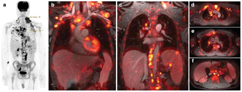
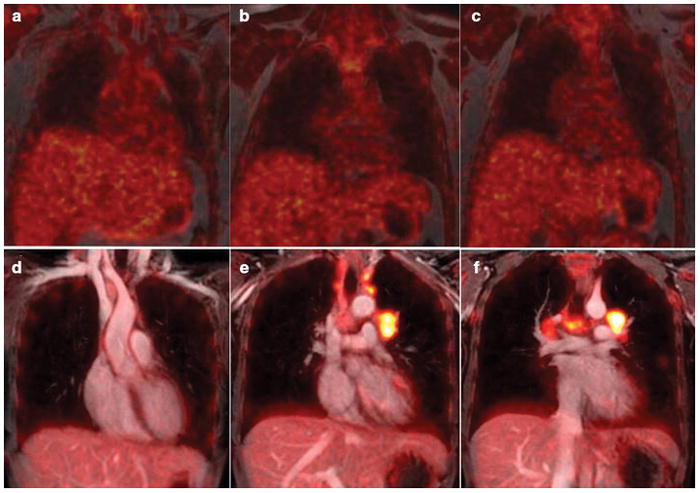
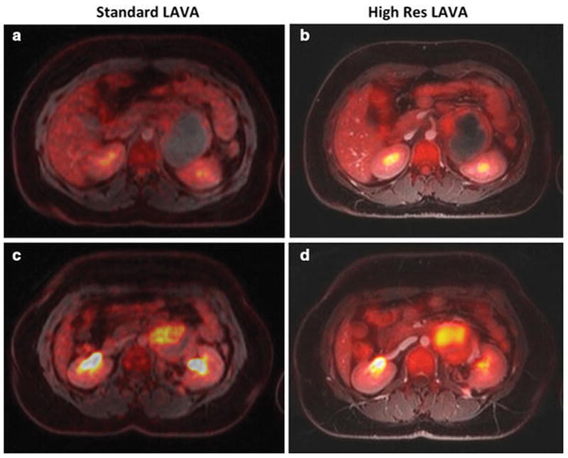
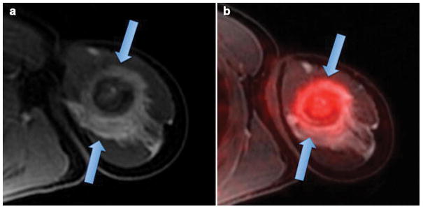
Recent findings: Since many primary tumors in children are evaluated with MRI and metastases are detected with PET/CT, integrated PET/MR can be a time-efficient and convenient solution for pediatric cancer staging. 18F-FDG PET/MR can assess primary tumors and the whole body in one imaging session, avoid repetitive anesthesia and reduce radiation exposure compared to 18F-FDG PET/CT. This article lists 10 action points, which might improve the clinical value of PET/MR for children with cancer. However, even if PET/MR proves valuable, it cannot enter mainstream applications if it is not accessible to the majority of pediatric cancer patients. Therefore, innovations are needed to make PET/MR scanners affordable and increase patient throughput.

Summary: PET/MR offers opportunities for more efficient, accurate and safe diagnoses of pediatric cancer patients. The impact on patient management and outcomes has to be substantiated by large-scale prospective clinical trials.

Keywords: Magnetic Resonance; PET/MR; Pediatric Cancer; Pediatric Lymphoma; Pediatric Sarcoma; Positron Emission Tomography.


Conflict of interest statement

Conflict of Interest Heike Daldrup-Link declares no potential conflicts of interest.

Figures

Figure 1
Figure 1. Whole Body 18F-FDG PET/MR of a patient with Hodgkin Lymphoma
(a) 18F-FDG PET maximum intensity projection shows FDG-avid disease of the neck, chest, axillaries, abdomen and pelvis. (b) Coronal 18F-FDG PET images superimposed on ferumoxytol-enhanced T1-weighted LAVA images clearly show the relation between vessels and multiple hypermetabolic lymph nodes. Three lesions in the spleen are also noted. (c) Axial integrated 18F-FDG PET/LAVA images provide diagnostic information similar to a PET/CT scan, but with improved soft tissue contrast.
Figure 2
Figure 2. Intravenous MR contrast agent administration improves soft tissue contrast of integrated PET/MR images
(a–c) Coronally reconstructed 18F-FDG PET images, superimposed on unenhanced T1-weighted LAVA images show limited soft tissue resolution. (d–f) Coronal 18F-FDG PET images, superimposed on ferumoxytol-enhanced T1-weighted LAVA images of a patient with Hodgkin lymphoma show improved soft tissue contrast: The ferumoxytol-enhanced 18F-FDG PET/MR images facilitate the delineation of mediastinal vessels and hypermetabolic lymph nodes. This is important, because current clinical protocols require accurate assessment of the tumor size and metabolic activity before, during and after therapy.
Figure 3
Figure 3. Spatial resolution on 18F-FDG PET/MR is important to render accurate diagnostic information in a young adult with retroperitoneal soft tissue sarcoma
(a,c) Axial 18F-FDG PET images, superimposed on ferumoxytol-enhanced T1-weighted LAVA images (TR/TE/alpha = 4.1ms/1.7ms/15), obtained for attenuation correction of 18F-FDG PET data. These images with a field of view (FOV) or 50 cm, a matrix of 256 x 128 pixels and a slice thickness of 5.2 mm provide limited anatomical resolution. For example, the superior mesenteric artery is not seen on these scans. (b,d) Axial 18F-FDG PET images, superimposed on ferumoxytol-enhanced T1-weighted LAVA images (TR/TE/alpha = 4.2ms/1.7ms/15), acquired for diagnostic purposes. These images with a field of view (FOV) or 48 cm, a matrix of 320 x 224 pixels and a slice thickness 3.4 mm provide improved tumor delineation from adjacent vessels and bowel. We conclude that current pulse sequences for AC correction do not have sufficient anatomical resolution to be used for diagnostic purposes.
Figure 4
Figure 4. Ferumoxytol shows differential enhancement of hypercellular hematopoietic marrow and tumor
(a) Coronal and axial 18F-FDG PET/MR in a young adult after chemotherapy shows diffusely hypermetabolic hematopoietic marrow, (b) axial diffusion-weighted MR images and coronal LAVA images in the same patient after intravenous injection of ferumoxytol show homogeneous iron oxide uptake in the bone marrow, as indicated by homogenous hypointense (dark) marrow signal. (c) Coronal ferumoxytol-enhanced T2-weighted fast spinecho scan of the lumbar spine and pelvis in an 11 year-old patient with biopsy-proven Hodgkin’s leukemia shows multiple focal tumor lesions in the bone marrow (arrows). The normal marrow shows negative (dark) ferumoxytol enhancement while focal tumors show little or no ferumoxytol uptake and stand out as hyperintense (bright) lesions.
Figure 5
Figure 5. 18F-FDG PET/MR shows differential enhancement of an osteosarcoma and peri-lesional edema
(a) Axial T1-weighted ferumoxytol-enhanced LAVA scan shows an ill defined, aggressive lesion in the proximal left humerus with extensive perilesional contrast-enhanced edema (arrows). (b) Integrated ferumoxytol-enhanced 18F-FDG PET/LAVA scan shows improved delineation of the 18F-FDG avid tumor (arrow) from enhancing peritumoral edema.

References

    1. Federman N, Feig SA. PET/CT in evaluating pediatric malignancies: a clinician’s perspective. Journal of nuclear medicine : official publication, Society of Nuclear Medicine. 2007;48(12):1920–2. doi: 10.2967/jnumed.107.046045. - DOI - PubMed
    1. Tatsumi M, Miller JH, Wahl RL. 18F-FDG PET/CT in evaluating non-CNS pediatric malignancies. Journal of nuclear medicine : official publication, Society of Nuclear Medicine. 2007;48(12):1923–31. doi: 10.2967/jnumed.107.044628. - DOI - PubMed
    1. Gerth HU, Juergens KU, Dirksen U, Gerss J, Schober O, Franzius C. Significant benefit of multimodal imaging: PET/CT compared with PET alone in staging and follow-up of patients with Ewing tumors. Journal of nuclear medicine : official publication, Society of Nuclear Medicine. 2007;48(12):1932–9. doi: 10.2967/jnumed.107.045286. - DOI - PubMed
    1. Kleis M, Daldrup-Link H, Matthay K, Goldsby R, Lu Y, Schuster T, et al. Diagnostic value of PET/CT for the staging and restaging of pediatric tumors. Eur J Nucl Med Mol Imaging. 2009;36(1):23–36. doi: 10.1007/s00259-008-0911-1. - DOI - PubMed
    1. Uslu L, Donig J, Link M, Rosenberg J, Quon A, Daldrup-Link HE. Value of 18F-FDG PET and PET/CT for evaluation of pediatric malignancies. J Nucl Med. 2015;56(2):274–86. doi: 10.2967/jnumed.114.146290. - DOI - PubMed

LinkOut - more resources