

Ror2 Signaling and Its Relevance in Breast Cancer Progression
- PMID: 28695110
- PMCID: PMC5483589
- DOI: 10.3389/fonc.2017.00135
Ror2 Signaling and Its Relevance in Breast Cancer Progression
Abstract
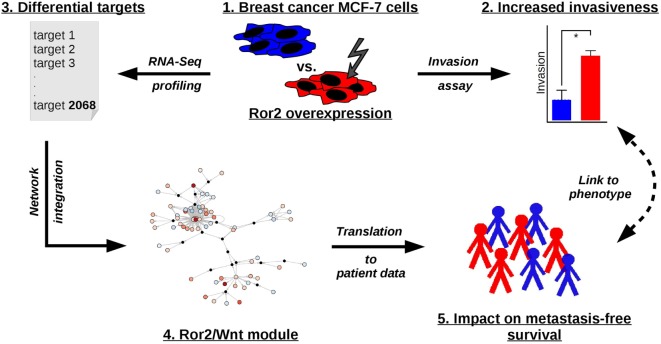
Breast cancer is a heterogeneous disease and has been classified into five molecular subtypes based on gene expression profiles. Signaling processes linked to different breast cancer molecular subtypes and different clinical outcomes are still poorly understood. Aberrant regulation of Wnt signaling has been implicated in breast cancer progression. In particular Ror1/2 receptors and several other members of the non-canonical Wnt signaling pathway were associated with aggressive breast cancer behavior. However, Wnt signals are mediated via multiple complex pathways, and it is clinically important to determine which particular Wnt cascades, including their domains and targets, are deregulated in poor prognosis breast cancer. To investigate activation and outcome of the Ror2-dependent non-canonical Wnt signaling pathway, we overexpressed the Ror2 receptor in MCF-7 and MDA-MB231 breast cancer cells, stimulated the cells with its ligand Wnt5a, and we knocked-down Ror1 in MDA-MB231 cells. We measured the invasive capacity of perturbed cells to assess phenotypic changes, and mRNA was profiled to quantify gene expression changes. Differentially expressed genes were integrated into a literature-based non-canonical Wnt signaling network. The results were further used in the analysis of an independent dataset of breast cancer patients with metastasis-free survival annotation. Overexpression of the Ror2 receptor, stimulation with Wnt5a, as well as the combination of both perturbations enhanced invasiveness of MCF-7 cells. The expression-responsive targets of Ror2 overexpression in MCF-7 induced a Ror2/Wnt module of the non-canonical Wnt signaling pathway. These targets alter regulation of other pathways involved in cell remodeling processing and cell metabolism. Furthermore, the genes of the Ror2/Wnt module were assessed as a gene signature in patient gene expression data and showed an association with clinical outcome. In summary, results of this study indicate a role of a newly defined Ror2/Wnt module in breast cancer progression and present a link between Ror2 expression and increased cell invasiveness.
Keywords: Ror2; Wnt signaling; breast cancer; metastasis; module; network integration.
Figures


Similar articles
-
WNT11/ROR2 signaling is associated with tumor invasion and poor survival in breast cancer.J Exp Clin Cancer Res. 2021 Dec 15;40(1):395. doi: 10.1186/s13046-021-02187-z. J Exp Clin Cancer Res. 2021. PMID: 34911552 Free PMC article.
-
The Wnt5a/Ror2 noncanonical signaling pathway inhibits canonical Wnt signaling in K562 cells.Int J Mol Med. 2011 Jan;27(1):63-9. doi: 10.3892/ijmm.2010.560. Epub 2010 Nov 10. Int J Mol Med. 2011. PMID: 21069266
-
Prognostic significance of Ror2 and Wnt5a expression in medulloblastoma.Brain Pathol. 2013 Jul;23(4):445-53. doi: 10.1111/bpa.12017. Epub 2013 Jan 25. Brain Pathol. 2013. PMID: 23278988 Free PMC article.
-
The Ror-Family Receptors in Development, Tissue Regeneration and Age-Related Disease.Front Cell Dev Biol. 2022 Apr 13;10:891763. doi: 10.3389/fcell.2022.891763. eCollection 2022. Front Cell Dev Biol. 2022. PMID: 35493090 Free PMC article. Review.
-
Insight into the role of Wnt5a-induced signaling in normal and cancer cells.Int Rev Cell Mol Biol. 2015;314:117-48. doi: 10.1016/bs.ircmb.2014.10.003. Epub 2014 Nov 18. Int Rev Cell Mol Biol. 2015. PMID: 25619716 Review.
Cited by
-
Opportunities and Challenges in the Development of Antibody-Drug Conjugate for Triple-Negative Breast Cancer: The Diverse Choices and Changing Needs.World J Oncol. 2024 Aug;15(4):527-542. doi: 10.14740/wjon1853. Epub 2024 Jul 5. World J Oncol. 2024. PMID: 38993251 Free PMC article. Review.
-
Explaining decisions of graph convolutional neural networks: patient-specific molecular subnetworks responsible for metastasis prediction in breast cancer.Genome Med. 2021 Mar 11;13(1):42. doi: 10.1186/s13073-021-00845-7. Genome Med. 2021. PMID: 33706810 Free PMC article.
-
ROR2 deficit may induce the tetralogy of Fallot via down-regulating of β-catenin/SOX3/HSPA6 in vitro and in vivo.J Cell Mol Med. 2023 Nov;27(22):3539-3552. doi: 10.1111/jcmm.17969. Epub 2023 Sep 25. J Cell Mol Med. 2023. PMID: 37749917 Free PMC article.
-
The WNT/ROR Pathway in Cancer: From Signaling to Therapeutic Intervention.Cells. 2021 Jan 12;10(1):142. doi: 10.3390/cells10010142. Cells. 2021. PMID: 33445713 Free PMC article. Review.
-
Monoclonal Antibodies and Antibody-drug Conjugates as Emerging Therapeutics for Breast Cancer Treatment.Curr Drug Deliv. 2024;21(7):993-1009. doi: 10.2174/1567201820666230731094258. Curr Drug Deliv. 2024. PMID: 37519200 Review.
References
LinkOut - more resources
Full Text Sources
Other Literature Sources
Molecular Biology Databases
Miscellaneous