. 2017 Jun 6;4(7):512-516.
doi: 10.1002/acn3.428. eCollection 2017 Jul.

Glioblastoma in natalizumab-treated multiple sclerosis patients

Affiliations

Glioblastoma in natalizumab-treated multiple sclerosis patients

Fabian Sierra Morales et al. Ann Clin Transl Neurol. .

Abstract

We present two natalizumab-treated multiple sclerosis patients who developed glioblastoma multiforme (GBM) with variable outcomes. One patient had an isocitrate dehydrogenase (IDH)-wildtype GBM with aggressive behavior, who declined treatment and died 13 weeks after symptoms onset. The other patient underwent resection of an IDH-mutant secondary GBM that arose from a previously diagnosed grade II astrocytoma. He is still alive 5 years after the diagnosis of GBM. JC virus was not detected in either case. Whether natalizumab played a role in the development of GBM in those patients deserves further investigation.


Figures
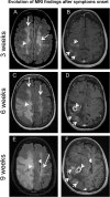
Figure 1
Figure 1
Progression of lesions in the right frontoparietal region on brain MRI. Axial fluid‐attenuated inversion recovery (FLAIR) (A, C, E) and postcontrast T1‐weighted images (B, D, F) demonstrate multiple irregular enhancing lesions (short arrows) with extensive surrounding edema. There is associated mass effect with leftward midline shift (arrow head). Multiple sclerosis lesions are present in the white matter of both hemispheres (long arrows).
Figure 2
Figure 2
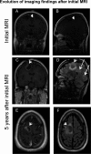
Initial MRI demonstrates a lesion (arrow head) without associate enhancement on coronal and sagittal postcontrast T1‐weighted images (A, B). Coronal and axial postcontrast T1‐weighted images (C, E) as well as sagittal, axial fluid‐attenuated inversion recovery (FLAIR) (D, F) demonstrate irregular enhancing lesion (short arrows) with surrounding edema. Multiple sclerosis lesions are visible in the subcortical white matter (long arrows).
Figure 3
Figure 3
Histology findings in patient #1 (A–B). (A) Glioblastoma multiforme, IDH1‐wildtype, WHO Grade IV with endothelial proliferation and necrosis (star) (H&E, 200x). (B) ATRX immunohistochemistry showing loss of nuclear staining in tumor cells (arrow), while retained nuclear staining in normal neurons (400×). Insert shows H&E of same area (H&E, 400×). Histology findings in patient #2 (C–F). (C) Diffuse astrocytoma, IDH1‐mutant, WHO Grade II from 2007 (H&E, 200×). (D) Glioblastoma multiforme with PNET‐like component, IDH1‐mutant, WHO Grade IV showing necrosis (star) and endothelial proliferation (arrowhead) (H&E 200×). (E) IDH1 R132H mutant staining showing strong cytoplasmic staining in the tumor cells and negative staining in the endothelial cells (400×). (F) ATRX immunostaining showing retained nuclear staining in the endothelial cells and loss of nuclear staining in the tumor cells (400×).

References

    1. Stupp R, Mason WP, Van den Bent MJ, et al. Radiotherapy plus concomitant and adjuvant temozolomide for glioblastoma. N Engl J Med 2005;352:987–996. - PubMed
    1. Compston A, Coles A. Multiple sclerosis. Lancet 2008;372:1502–1517. - PubMed
    1. Polman CH, O'Connor PW, Havrdova E, et al. A randomized, placebo‐controlled trial of natalizumab for relapsing multiple sclerosis. N Engl J Med 2006;354:899–910. - PubMed
    1. Rudick RA, Stuart WH, Calabresi PA, et al. Natalizumab plus interferon beta‐1a for relapsing multiple sclerosis. N Engl J Med 2006;354:911–923. - PubMed
    1. Law JY, Kim DW, Sturgis A, Naina H. Non‐hodgkin lymphoma of the stomach in a patient treated with natalizumab. Clin Med Insights Oncol 2015;1:61–63. - PMC - PubMed