

Thyroid Nodule Classification in Ultrasound Images by Fine-Tuning Deep Convolutional Neural Network
- PMID: 28695342
- PMCID: PMC5537102
- DOI: 10.1007/s10278-017-9997-y
Thyroid Nodule Classification in Ultrasound Images by Fine-Tuning Deep Convolutional Neural Network
Abstract
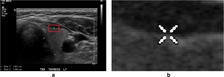
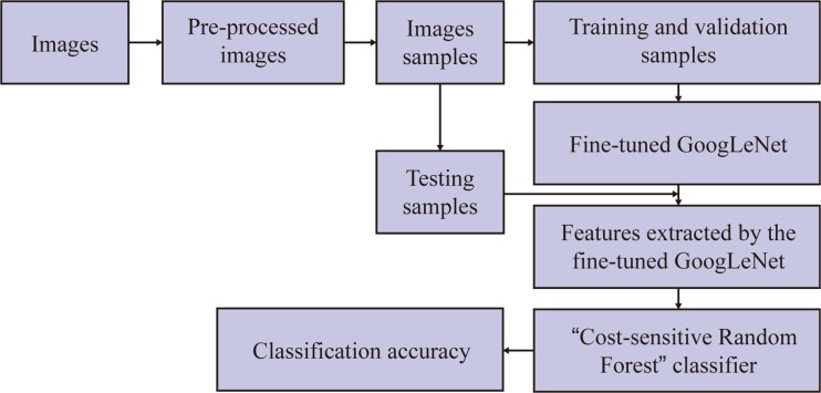
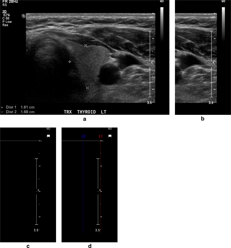
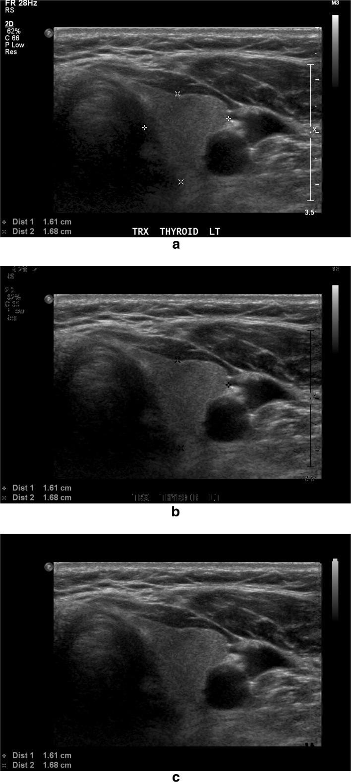
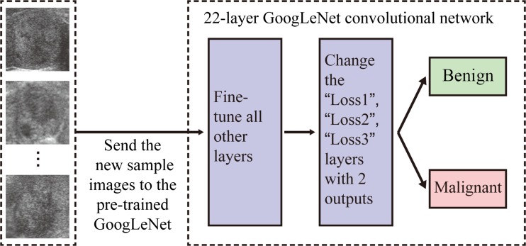
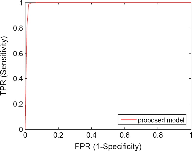
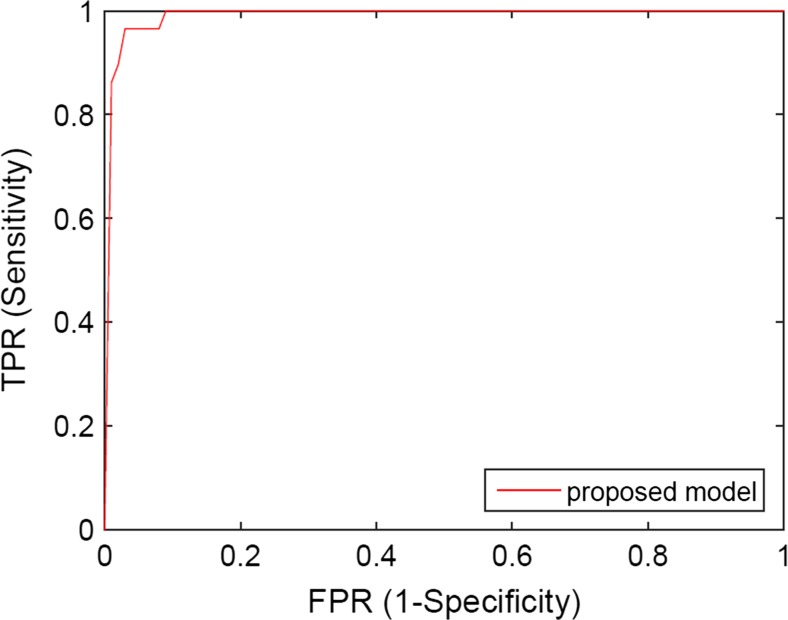
With many thyroid nodules being incidentally detected, it is important to identify as many malignant nodules as possible while excluding those that are highly likely to be benign from fine needle aspiration (FNA) biopsies or surgeries. This paper presents a computer-aided diagnosis (CAD) system for classifying thyroid nodules in ultrasound images. We use deep learning approach to extract features from thyroid ultrasound images. Ultrasound images are pre-processed to calibrate their scale and remove the artifacts. A pre-trained GoogLeNet model is then fine-tuned using the pre-processed image samples which leads to superior feature extraction. The extracted features of the thyroid ultrasound images are sent to a Cost-sensitive Random Forest classifier to classify the images into "malignant" and "benign" cases. The experimental results show the proposed fine-tuned GoogLeNet model achieves excellent classification performance, attaining 98.29% classification accuracy, 99.10% sensitivity and 93.90% specificity for the images in an open access database (Pedraza et al. 16), while 96.34% classification accuracy, 86% sensitivity and 99% specificity for the images in our local health region database.
Keywords: Computer vision; Convolutional neural network; Deep learning; Fine-tuning; Machine learning; Thyroid nodules; Ultrasonography.
Conflict of interest statement
Funding
This research was funded through a Collaborative Innovation Development Grant from the Saskatchewan Health Research Foundation.
Figures
References
-
- Diana Gaitini, Rhodri M Evans, and Gordana Ivanac, Chapter 16: thyroid ultrasound. EFSUMB Course Book, 2011.
-
- Chen S-J, Chang C-Y, Chang K-Y, Tzeng J-E, Chen Y-T, Lin C-W, Hsu W-C, Wei C-K. Classification of the thyroid nodules based on characteristic sonographic textural feature and correlated histopathology using hierarchical support vector machines. Ultrasound Med Biol. 2010;36(12):2018–2026. doi: 10.1016/j.ultrasmedbio.2010.08.019. - DOI - PubMed
-
- Chang C-Y, Liu H-Y, Tseng C-H, Shih S-R. Computer-aided diagnosis for thyroid Graves’ disease in ultrasound images. Biomed Eng: Appl Basis Commun. 2010;22(02):91–99.
MeSH terms
LinkOut - more resources
Full Text Sources
Other Literature Sources
Medical
Miscellaneous
