. 2017:1608:445-473.
doi: 10.1007/978-1-4939-6993-7_28.

Identifying and Validating Tankyrase Binders and Substrates: A Candidate Approach

Affiliations

Identifying and Validating Tankyrase Binders and Substrates: A Candidate Approach

Katie Pollock et al. Methods Mol Biol. 2017.

Abstract

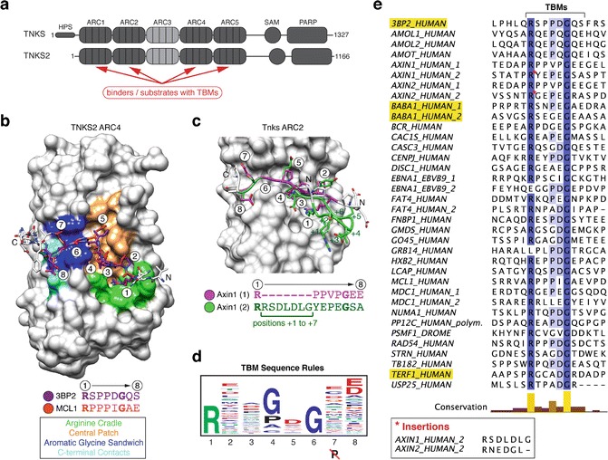
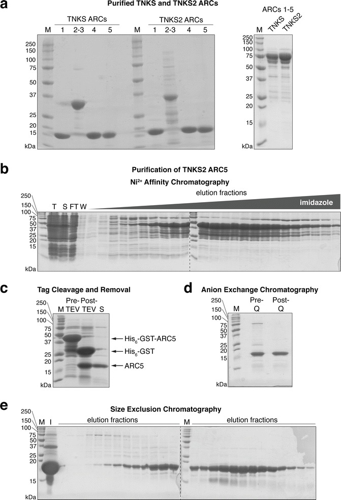
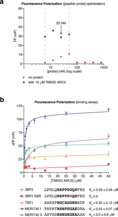
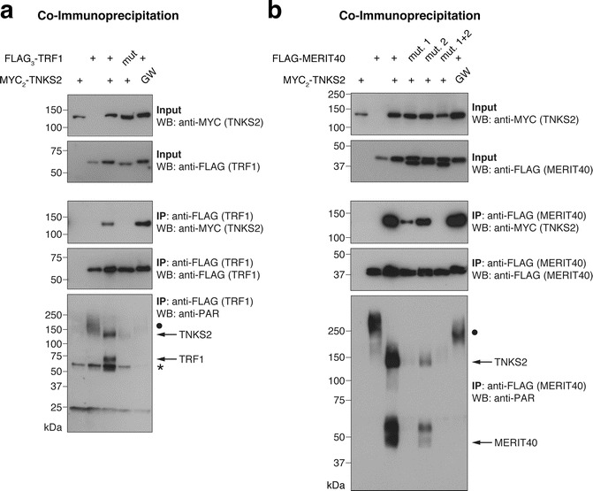
The poly(ADP-ribose)polymerase (PARP) enzyme tankyrase (TNKS/ARTD5, TNKS2/ARTD6) uses its ankyrin repeat clusters (ARCs) to recognize degenerate peptide motifs in a wide range of proteins, thereby recruiting such proteins and their complexes for scaffolding and/or poly(ADP-ribosyl)ation. Here, we provide guidance for predicting putative tankyrase-binding motifs, based on the previously delineated peptide sequence rules and existing structural information. We present a general method for the expression and purification of tankyrase ARCs from Escherichia coli and outline a fluorescence polarization assay to quantitatively assess direct ARC-TBM peptide interactions. We provide a basic protocol for evaluating binding and poly(ADP-ribosyl)ation of full-length candidate interacting proteins by full-length tankyrase in mammalian cells.

Keywords: Enzyme–substrate relationships; Fluorescence polarization (FP); PARP; Poly(ADP-ribosyl)ation; Protein expression; Protein purification; Protein-protein interactions; Structural biology; Tankyrase; Tankyrase-binding peptide motif.


Figures

Fig. 1
Fig. 1
Substrate binding by Tankyrase. (a) Domain organization of human tankyrase and tankyrase 2 (modified from [31]). (b) and (c) Examples for ARC–TBM interactions studied by X-ray crystallography. (b) Human TNKS2 ARC4 is shown in surface representation with bound TBM peptidesTankyrase Binders from 3BP23BP2 (SH3BP2, SH3 domain-binding protein 2) and MCL1 shown in stick representation with the core TBM octapeptide colored purple and orange, respectively, and by heteroatom. TBM amino acid positions (1–8) and sequences shown. The figure was generated by superimposing the ARCs of the ARC4-3BP2 and ARC4-MCL1 crystal structures (PDB accession codes 3TWR and 3TWU, respectively) onto each other and showing ARC4 of the former [7]. The colored surface areas represent different contact areas, as indicated, that mediate binding of the TBM peptides (Modified from [7] with permission from Elsevier/Cell Press). (c) ARC2 (from ARC2–3) of murine Tnks bound by the N-terminus of murine Axin1 (PDB accession code 3UTM), which contains two TBMs [11]. Each TBM binds one copy each of ARC2 in a dimeric ARC2-3 assembly. The figure was generated by superimposing the two ARC2-3 copies onto each other; the surface of ARC2 bound by the first TBM is shown. TBMs are shown and labeled as in (b). The first TBM, shown in magenta, consists of a continuous stretch of eight amino acids. In the second TBM, shown in green, the Arg at position 1 is followed by a seven-amino-acid insertion (positions +1 to +7), as indicated in the sequences shown. The peptide insertion forms a loop. (d) TBM sequence rulesSequence consensus/rules represented by a sequence logo. (Reprinted from [7] with permission from Elsevier/Cell Press.) (e) Sequence alignment of known example TBMs ([7] and references therein, [, , , –39]), colored by identity with conservation graph, generated with ClustalX and Jalview [40, 41]. UniProtUniProt database IDs are indicated [24]. The asterisk indicates insertion sequences in AXIN1 and AXIN2. The TBMs of 3BP2, TRF1 (TERF1_HUMAN), and MERIT40 (BABA1_HUMAN), studied as model TBMs here, are highlighted
Fig. 2
Fig. 2
Purification of tankyrase ARCs. (a) 5 μg purified TNKSTankyrase Binders and TNKS2 ARCs were resolved on 15% Tris–glycine polyacrylamide gels for SDS-PAGE and the gels stained with Coomassie. M, marker. See Table 1 for construct boundaries. (b)-(e) Step-by-step purification of a representative ARC, TNKS2 ARC5. (b) Ni2+ affinity purification of His6-GST-TNKS2 ARC5 fusion protein. T, total lysate; S, soluble lysate; FT, flow-through; W, wash. (c) Removal of the His6-GSTGST tag His6-GST tag affinity tag by TEV cleavage and a second Ni2+ affinity purification step. Pre-TEV, protein before TEV cleavage; Post-TEV, protein after TEV cleavage; S, pooled protein after subtraction of affinity tag. (d) Anion exchange chromatographyAnion exchange chromatography (Mono Q column). Pre-Q, protein before anion exchange chromatography; Post-Q, protein after anion exchange chromatography. (e) Size exclusion chromatography. M, marker; I, input
Fig. 3
Fig. 3
Assessing ARC–TBM peptide interactionsARC-peptide interactions by fluorescence polarizationFluorescence. (a) Optimization of the peptide probe (3BP2) concentration for the FP binding assay. Data are from one experiment performed in technical duplicate with mean FP values plotted. The dashed vertical lines indicate the suitable peptide probe concentration range. 25 nM was chosen for the subsequent assays. (b) FP binding assays for the TBM peptides from TRF1, MERIT40, and 3BP2 (WT positive control and G6R negative control). Peptide sequences (octapeptide in bold) and affinities for TNKS2 ARC5 are indicated. n = 3 separate experiments; error bars, SEM. The error values for the dissociation constants correspond to the standard error of the fit in nonlinear regression. n.d., not determined (no binding curve)
Fig. 4
Fig. 4
Assessing Tankyrase binding and PARylation by tankyrase in the full-length protein context. (a) FLAG3-TRF1, either in its wild-typeTankyrase Binders form or as a G18R TBM mutant (“mut.”), was co-expressed with the indicated MYC2-TNKS2 constructs, either in wild-type form or as a G1032W PARP-inactive mutant (“GW”) [31]. FLAG-TRF1 was immunoprecipitated and input and immunoprecipitate (IP) samples analyzed by SDS-PAGE and Western blotting as indicated. “•” in the anti-PAR blot labels a high-molecular-weight PARylated species that appears to be antagonized by MYC2-TNKS2 overexpression. “*” in the anti-PAR blot denotes a nonspecific band. (b) Same analysis as in (a) with FLAG-MERIT40, either in wild-type form or as a G33R (“mut. 1”), G53R (“mut. 2”), or GG33/53RR (“mut. 1 + 2”) TBM mutant. MERIT40 appears as a doublet, most likely reflecting differentially phosphorylated species [32]. See Note 22 on attributing PAR signals to candidate proteins

References

    1. Gibson BA, Kraus WL. New insights into the molecular and cellular functions of poly(ADP-ribose) and PARPs. Nat Rev Mol Cell Biol. 2012;13:411–424. doi: 10.1038/nrm3376. - DOI - PubMed
    1. Teloni F, Altmeyer M. Readers of poly(ADP-ribose): designed to be fit for purpose. Nucleic Acids Res. 2016;44:993–1006. doi: 10.1093/nar/gkv1383. - DOI - PMC - PubMed
    1. Vyas S, Chesarone-Cataldo M, Todorova T, et al. A systematic analysis of the PARP protein family identifies new functions critical for cell physiology. Nat Commun. 2013;4:2240. doi: 10.1038/ncomms3240. - DOI - PMC - PubMed
    1. Hsiao SJ, Smith S. Tankyrase function at telomeres, spindle poles, and beyond. Biochimie. 2008;90:83–92. doi: 10.1016/j.biochi.2007.07.012. - DOI - PubMed
    1. Haikarainen T, Krauss S, Lehtio L. Tankyrases: structure, function and therapeutic implications in cancer. Curr Pharm Des. 2014;20:6472–6488. doi: 10.2174/1381612820666140630101525. - DOI - PMC - PubMed

Publication types

LinkOut - more resources