. 2017:1608:475-513.
doi: 10.1007/978-1-4939-6993-7_29.

Computational and Experimental Studies of ADP-Ribosylation

Affiliations

Computational and Experimental Studies of ADP-Ribosylation

Robert G Hammond et al. Methods Mol Biol. 2017.

Abstract

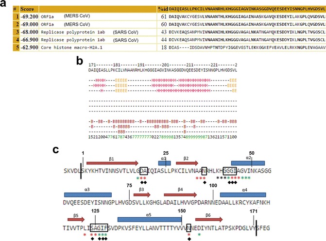
The macrodomains are a multifunctional protein family that function as receptors and enzymes acting on poly(ADP-ribose), ADP-ribosylated proteins, and other metabolites of nicotinamide adenine dinucleotide (NAD+). Several new functions for macrodomains, such as nucleic acid binding and protein-protein interaction, have recently been identified in this family. Here, we discuss methods for the identification of new macrodomains in viruses and the prediction of their function. This is followed by the expression and purification of these proteins following overexpression in bacterial cells and confirmation of folding and function using biophysical methods.

Keywords: Bioinformatics; Circular dichroism; Coronavirus; Docking; Expression; Macrodomain; NMR spectroscopy; Purification; STD-NMR.


Figures

Fig. 1
Fig. 1
(a) Result of a FFAS database search using the bat CoV HKU4 nsp3 protein sequence. The top five results, their sequence identity, FFAS score and protein names are displayed. The Protein Data Bank served as the database for this search . The full result is truncated for legibility and virus names were added for clarity. (b) Jpred output from HKU4 query: β-sheet—E, α-helix—H, buried amino acids—B. Confidence scores are listed at the bottom: (0) lowest, (9) highest. (c) Predicted macrodomain in HKU4. Predicted secondary structure, βαβααββαβαβ, is shown above the sequence. Vertical lines and numbering indicate the start and endpoints of the predicted domain. Conserved residues of viral macrodomains that are directly involved in ADP-ribose recognition are boxed in black. Residues predicted to be involved in ADP-ribose recognition via output of the BioLiP database and COACH program are shown with green and black stars, respectively. Red stars indicate residues predicted to be involved in ADP-ribose recognition by both BioLiP and COACH. Black diamonds indicate residues that are conserved with respect to residues involved in interactions with the ligand, in the SARS-CoV macrodomain complex identified by Glide docking (Fig. 2a)
Fig. 2
Fig. 2
(a) Docked structure from Glide of the complex between ADP-ribose and the SARS-CoV macrodomain. ADP-ribose is shown as a stick model and colored by elements: white, hydrogen; green, carbon; red, oxygen; blue, nitrogen; purple, phosphorus. The protein is shown in a ribbon representation , with selected interacting residues in the binding cavity shown as ball-and-stick and labeled. For comparison, the X-ray crystallographic position of the ligand (PDB: 2FAV [59]) is shown as a wire-frame model in dark green. The two ligands exhibit a similar binding mode (RMSD = 0.74 Å). (b) Docked complex from ZDOCK between the SARS-CoV unique domain M (PDB: 2JZE [90]) and I14–Tel23, an antiparallel basket G-quadruplex from human telomeric DNA (PDB: 2KKA) [91]. The protein is shown as a ribbon model in green, and interacting residues of SUD-M are labeled and shown as stick models in red. The quadruplex is shown in cartoon mode in blue; interacting bases of the quadruplex are shown as cartoon ring mode in magenta. Predicted hydrogen bonds between the two molecules are shown as yellow dashed lines
Fig. 3
Fig. 3
(a) Chromatogram of the HKU4 putative macrodomain size-exclusion chromatography purification. The protein elution is indicated by the major peak. UV detection at 254 nm and 280 nm is indicated by the red and blue traces. (b) Elution of protein (18.4 kDa) monitored by SDS-PAGE (4–12% Bis-Tris gel) of selected fractions collected from the chromatography run in (a)

References

    1. de Groot RJ, Baker SC, Baric RS, Brown CS, Drosten C, Enjuanes L, Fouchier RA, Galiano M, Gorbalenya AE, Memish ZA, Perlman S, Poon LL, Snijder EJ, Stephens GM, Woo PC, Zaki AM, Zambon M, Ziebuhr J. Middle East respiratory syndrome coronavirus (MERS-CoV): announcement of the Coronavirus Study Group. J Virol. 2013;87(14):7790–7792. doi: 10.1128/JVI.01244-13. - DOI - PMC - PubMed
    1. Zumla A, Hui DS, Perlman S. Middle East respiratory syndrome. Lancet. 2015;386(9997):995–1007. doi: 10.1016/S0140-6736(15)60454-8. - DOI - PMC - PubMed
    1. Peiris JS, Guan Y, Yuen KY. Severe acute respiratory syndrome. Nat Med. 2004;10(12 Suppl):S88–S97. doi: 10.1038/nm1143. - DOI - PMC - PubMed
    1. Fehr AR, Perlman S. Coronaviruses: an overview of their replication and pathogenesis. Methods Mol Biol. 2015;1282:1–23. doi: 10.1007/978-1-4939-2438-7_1. - DOI - PMC - PubMed
    1. Frieman M, Baric R (2008) Mechanisms of severe acute respiratory syndrome pathogenesis and innate immunomodulation. Microbiol Mol Biol Rev 72(4):672–685, Table of Contents. doi:10.1128/MMBR.00015-08 - PMC - PubMed

Publication types

MeSH terms

LinkOut - more resources