

Comment
doi: 10.1038/cr.2017.92.
Epub 2017 Jul 11.
Coding GPCR-G protein specificity
Affiliations
- PMID: 28695889
- PMCID: PMC5630685
- DOI: 10.1038/cr.2017.92
Item in Clipboard
Comment
Coding GPCR-G protein specificity
Cell Res.
2017 Oct.
Abstract
G protein-coupled receptors (GPCRs) are ubiquitous gatekeepers of cellular response and signal predominantly by recruitment and activation of G proteins. In a recent paper in Nature, Flock et al. use large-scale bioinformatics to build a model of GPCR-G protein selectivity and an interactive database to interrogate potential receptor-G protein interactions.
Figures
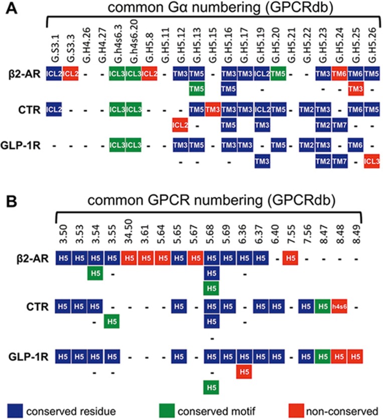
Selectivity determinants from GPCRdb were mapped onto Gαsβγ:β2AR (3sn6), Gαsβγ:CTR (5uz7) and Gαsβγ:GLP-1R (5vai), and residues within ∼4 Å and showing an appropriate interaction were manually annotated (other residues are not shown). In A, residues from the Gα interface are mapped against the 3 receptors. Gα positions are labeled using the common Gα numbering system (GPCRdb) where S3 is β-strand 3, h4s6 is the loop between helix 4 and β-strand 6 and H5 is helix 5 (all in the Ras-like domain). These are shown against interacting residues in the 3 GPCRs; in blue are GPCR residues that have corresponding positions (according to GPCRdb), green are GPCR residues in corresponding secondary structures but at alternative positions and red illustrates unique GPCR residues. In B, residues from the G protein-coupling pocket are mapped against GPCRs (only interacting residues are shown); the color scheme is the same as A but applies to Gαs. Pocket residues are numbered according to GPCRdb in which the first (single) number indicates the transmembrane helix or, in the case of 2 numbers, the loop connecting 2 helices. The number to the right of the decimal indicates the position of this residue relative to the most conserved helix residue, which is given the value of 50.
Comment on
-
Selectivity determinants of GPCR-G-protein binding.Nature. 2017 May 18;545(7654):317-322. doi: 10.1038/nature22070. Epub 2017 May 10. Nature. 2017. PMID: 28489817 Free PMC article.
References
Publication types
MeSH terms
Substances
LinkOut - more resources
Full Text Sources
Other Literature Sources
