

Editorial
doi: 10.1038/cdd.2017.111.
CDPs: caspase-dependent non-lethal cellular processes
Affiliations
- PMID: 28695898
- PMCID: PMC5520448
- DOI: 10.1038/cdd.2017.111
Item in Clipboard
Editorial
CDPs: caspase-dependent non-lethal cellular processes
Cell Death Differ.
2017 Aug.
No abstract available
Conflict of interest statement
The authors declare no conflict of interest.
Figures
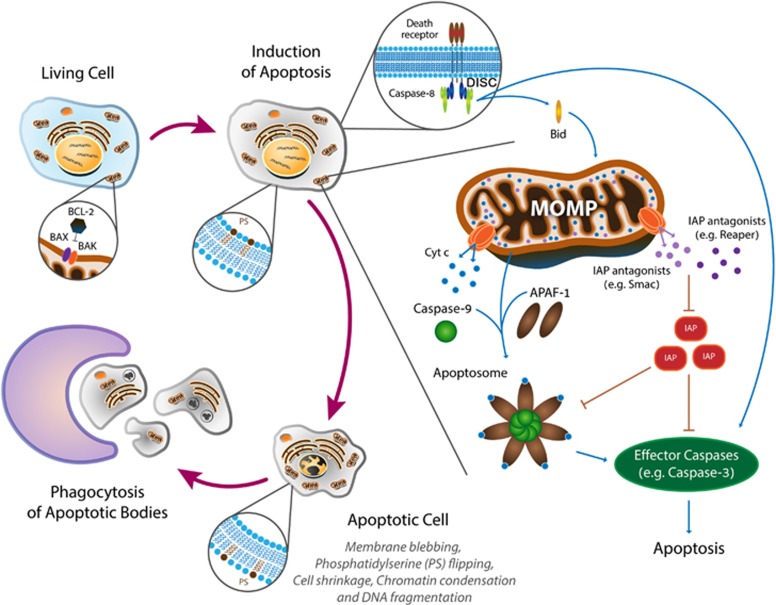
Schematic diagram of the conserved biochemical machinery and the corresponding morphological changes during apoptosis
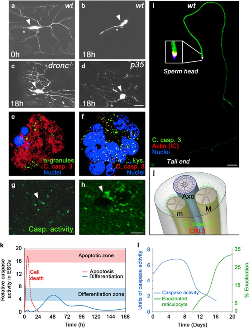
Different CDP paradigms. (a–d), Dendritic pruning in ddaC neurons in the Drosophila pupa requires caspase activity. Eighteen hours into metamorphosis all the ddaC neuron’s dendrites are eliminated (a,b). In flies mutant for the initiator caspase (dronc−/−) pruning is completely blocked (c). However, overexpression of the effector caspase inhibitor protein p35 only attenuates the elimination of the severed dendrites, suggesting an additional role for the initiator caspase unrelated to its role in the activation of the effector caspases (d). Arrowheads indicate the neuronal cell bodies, while asterisks indicate the unpruned major axon. Scale bar, 20 μm. (e,f), Activated caspases are confined to cytoplasmic granules of unknown nature during platelets formation in maturing megakaryocytes (MKs). Proplatelet-displaying MKs induced to undergo apoptosis by staurosporine display diffused active caspase-3 throughout the cytoplasm (e). In contrast, active caspase-3 was confined to cytoplasmic granules that do not co-localize with lysosomes (Lys.) or α-granules in these maturing MKs (not shown; f). (g,h), Dendritic spines in the auditory forebrain of adult zebra finches during song habituation training. Sections from the auditory forebrain taken from a bird hearing only silence (g) or a bird immediately after listening to ten novel songs (h). Arrowheads indicate the areas of the dendritic spines where a transient increase in the density of caspase-3 active sites is detected after song habituation training. Scale bar, 4 μm. (i), Drosophila spermatids at the onset of the individualization process. A cyst of 64-spermatid bundle displaying caspase activation at a graded pattern descending from sperm head to tail end. Note, that the cytoplasmic contents together with the active caspases are gradually removed at the same direction as that of caspase activation, such that the regions that are the first to individualize encounter the highest levels of active caspases but for the shortest time. Scale bar, 50 μm. (j), An illustration of a cross section through an elongated spermatid depicting the axoneme (Axo; blue) attached to the two mitochondrial derivatives (M and m; gold). The CRL3 complex is expressed throughout the cytoplasm (red dots). However, once CRL3 binds to the mitochondrial surface (through a protein called A-Sβ), it gets activated, and in turn activates effector caspases in the vicinity of the mitochondria (green dots), which then diffuse in the cytoplasm. (k), Changes in the intensity and duration of caspase activity can determine cell death versus cell differentiation of mESCs into cardiomyocytes. Caspase-3/7 activity was monitored in mESC lysates at different time points showing a rapid and intense elevation in caspase activity following a death signal (red line), and delayed and reduced caspase activity levels associated with differentiation (blue line). (l), Casapase-3 activity precedes enucleation of red blood cells. The graphs describe erythropoiesis induced in culture. A peak in caspase-3 is recorded several days before the enucleation process begins. The figures were adopted with permission from the following papers: (a–d) from ref. ; (e,f) from ref. ; (g,h) from ref. ; (i) from ref. ; (j) was modified from ref. ; (k) is a different presentation of the data in Figure 5 from ref. ; (l) is a combined presentation of the data in Figures 1 and 3 from ref.
References
Publication types
MeSH terms
Substances
LinkOut - more resources
Full Text Sources
Other Literature Sources
Molecular Biology Databases
