. 2018 Feb;36(2):739-750.
doi: 10.1002/jor.23651. Epub 2017 Jul 31.

Mitochondrial dysfunction is an acute response of articular chondrocytes to mechanical injury

Affiliations

Mitochondrial dysfunction is an acute response of articular chondrocytes to mechanical injury

Michelle L Delco et al. J Orthop Res. 2018 Feb.

Abstract

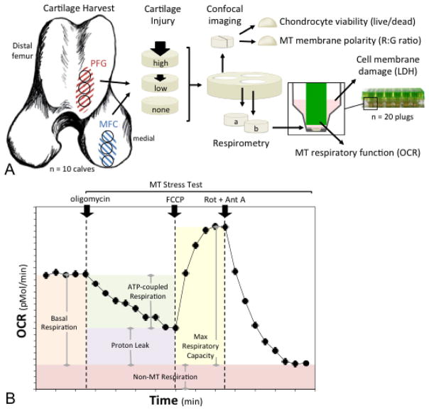
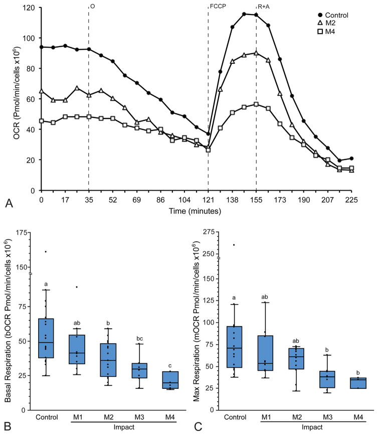
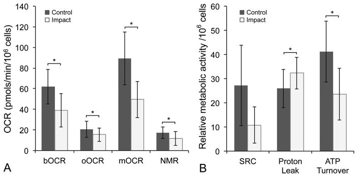
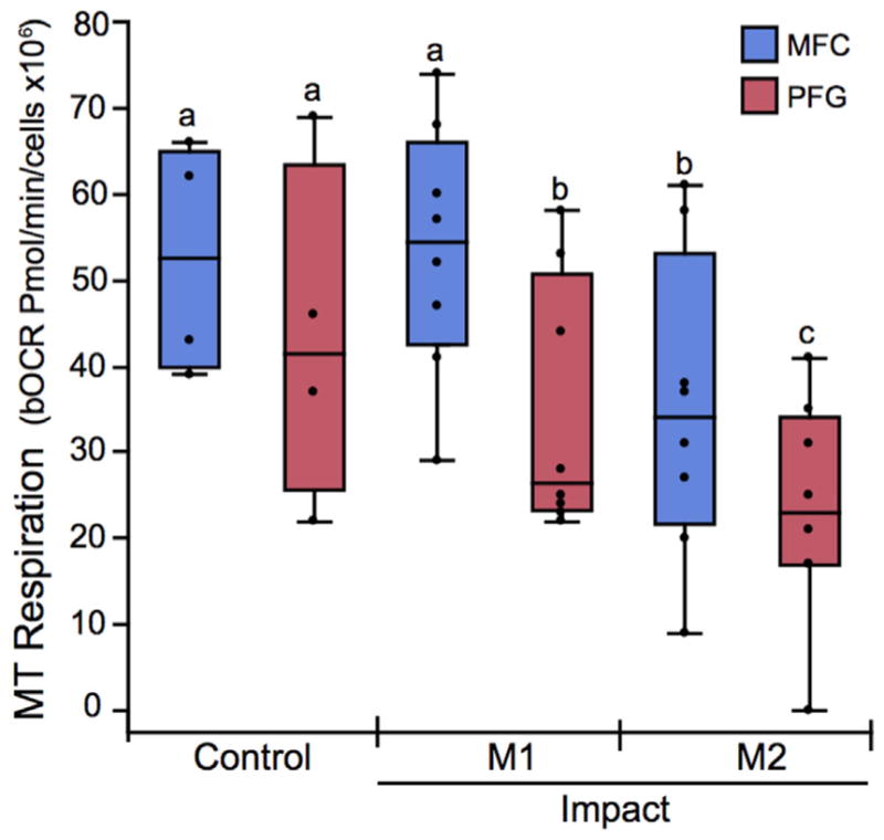
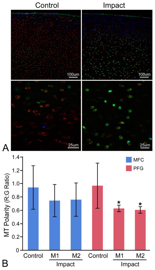
Mitochondrial (MT) dysfunction is known to occur in chondrocytes isolated from end-stage osteoarthritis (OA) patients, but the role of MT dysfunction in the initiation and early pathogenesis of post-traumatic OA (PTOA) remains unclear. The objective of this study was to investigate chondrocyte MT function immediately following mechanical injury in cartilage, and to determine if the response to injury differed between a weight bearing region (medial femoral condyle; MFC) and a non-weight bearing region (distal patellofemoral groove; PFG) of the same joint. Cartilage was harvested from the MFC and PFG of 10 neonatal bovids, and subjected to injurious compression at varying magnitudes (5-17 MPa, 5-34 GPa/s) using a rapid single-impact model. Chondrocyte MT respiratory function, MT membrane polarity, chondrocyte viability, and cell membrane damage were assessed in situ. Cartilage impact resulted in MT depolarization and impaired MT respiratory function within 2 h of injury. Cartilage from a non-weight bearing region of the joint (PFG) was more sensitive to impact-induced MT dysfunction and chondrocyte death than cartilage from a weight-bearing surface (MFC). Our findings suggest that MT dysfunction is an acute response of chondrocytes to cartilage injury, and that MT may play a key mechanobiological role in the initiation and early pathogenesis of PTOA.

Clinical significance: Direct therapeutic targeting of MT function in the early post-injury time frame may provide a strategy to block perpetuation of tissue damage and prevent the development of PTOA. © 2017 Orthopaedic Research Society. Published by Wiley Periodicals, Inc. J Orthop Res 36:739-750, 2018.

Keywords: cartilage; mechanobiology; mitochondria; osteoarthritis; posttraumatic.


Figures

Figure 1
Figure 1
Experimental design and methods. (A) Cartilage explants were harvested from the medial femoral condyle (MFC) for the first set of experiments, and from two sites for the second set of experiments; the MFC and the distal patellofemoral groove (PFG). One explant from each region was impacted at a higher impact magnitude, one was impacted at a lower magnitude, and one served as an un-impacted control. Explants were then divided for use in several assays; chondrocyte viability was quantified using live/dead staining, mitochondrial (MT) membrane polarity was determined as red to green fluorescent intensity (R:G) ratio on confocal imaging and MT respiratory function was assessed via microrespirometry. Cell membrane damage was assessed by measuring lactate dehydrogenase (LDH) activity in cartilage conditioned media. (B) MT respiratory function was quantified by measuring oxygen consumption rate (OCR) over time. After basal OCR was determined, a MT stress test was performed by sequential addition of (i) oligomycin, an ATP synthase inhibitor; (ii) FCCP, a proton circuit uncoupler; and (iii) rotenone (Rot) and antimycin A (Ant A), inhibitors of MT complexes I and III. Parameters of respiratory control were calculated as depicted by the shaded areas under the curve.
Figure 2
Figure 2
Respirometry reveals acute impact-induced respiratory impairment. Cartilage from the medial femoral condyle was impacted at various magnitudes (M1–M4) and MT respiration was quantified by measuring oxygen consumption rate (OCR), then normalizing data to live cell number for each explant. (A) Representative curves for OCR versus time for control, low impact (M2), and high impact (M4) groups demonstrate differences in MT respiration between groups. Note that oligomycin-inhibited respiration (oOCR) does not reach steady state (121 min), but Rot + AA inhibited respiration does (225 min). (B) Basal OCR (bOCR) and (C) max respiration (mOCR) decreased with increasing impact magnitude (M1–M4). Groups that do not share a letter are significantly different at p <0.05. Box and whisker plots represent data quartiles.
Figure 3
Figure 3
Mitochondrial dysfunction occurs after cartilage injury. (A) Values for bOCR, oOCR, mOCR, and non-MT respiration (NMR) and (B) spare respiratory capacity (SRC), proton leak (as a % of bOCR), and ATP turnover in injured versus control samples. Error bars = ±s.d.; *p <0.05.
Figure 4
Figure 4
Impact-induced chondrocyte death differs by location within the joint. (A) Chondrocyte death is correlated with impact magnitude. Cell death was positively correlated with peak impact stress for both the MFC (r2 = 0.70, p <0.0001) and the PFG (r2 = 0.79, p <0.001). Data presented is for all impacts performed throughout the study. (B) Chondrocytes from the patellofemoral groove (PFG) were more sensitive to impact-induced cell death than the medial femoral condyle (MFC). At lower impact magnitudes (M1) MFC viability was not affected. Groups that do not share a letter are significantly different at p <0.05. Error bars, ±s.d. (C and D) Representative images of PFG cartilage stained for live cells (green) with calcein AM and dead cells (red) with ethidium homodimer and imaged in cross-section using confocal microscopy. The articular surface is toward the top of the images. (C) Un-impacted control cartilage had less dead (red) staining than (D) lower impacted (M1) and (E) higher impacted (M2) in PFG explants.
Figure 5
Figure 5
The patellofemoral groove (PFG) is more sensitive to impact-induced respiratory impairment than the medial femoral condyle (MFC). The basal oxygen consumption rate (bOCR) of viable chondrocytes was significantly lower in PFG cartilage (red box and whisker quartile plots) impacted at the lowest (M1) and higher (M2) magnitudes compared to un-injured control cartilage, whereas in MFC cartilage (blue plots), bOCR is only affected at the higher impact magnitude (M2). Groups that do not share a letter are significantly different at p <0.05.
Figure 6
Figure 6
The patellofemoral groove (PFG) is more sensitive to impact-induced MT depolarization than the medial femoral condyle (MFC). (A) Representative confocal images of control and impacted (M2) PFG explants stained for MT polarity at low (top) and high (bottom) magnification. Cartilage is stained with Mitotracker Green (green; all MT), tetramethylrhodamine methyl ester perchlorate (red; polarized/functional MT), and Hoechst 33342 (blue; nuclear counterstain, higher affinity for cells with compromised cell membranes). Red:green fluorescent intensity ratios were calculated on an image-wide basis in multiple low magnification z-stacks for each explant (top) using a custom ImageJ macro. This high-throughput technique was validated by manually drawing ROIs around individual cells at higher magnification (bottom) to calculate R:G ratios on a single-cell basis. (B) MT depolarization occurred in PFG cartilage from both the lower (M1) and higher impact (M2) groups compared to PFG controls. Significant differences were not detected between impact groups from the MFC. Asterisks denote a significant difference compared to control at p <0.05. Error bars = ±s.d.

References

    1. Buckwalter JA. The role of mechanical forces in the initiation and progression of osteoarthritis. HSS Jrnl. 2012;8:37–38. - PMC - PubMed
    1. Kramer WC, Hendricks KJ, Wang J. Pathogenetic mechanisms of posttraumatic osteoarthritis: opportunities for early intervention. Int J Clin Exp Med. 2011;4:285–298. - PMC - PubMed
    1. Cheng DS, Visco CJ. Pharmaceutical therapy for osteoarthritis. PM&R. 2012;4:S82–S88. - PubMed
    1. Anderson DD, Chubinskaya S, Guilak F, et al. Post-traumatic osteoarthritis: improved understanding and opportunities for early intervention. J Orthop Res. 2011;29:802–809. - PMC - PubMed
    1. Chubinskaya S, Wimmer MA. Key pathways to prevent posttraumatic arthritis for future molecule-based therapy. Cartilage. 2013;4:13S–21S. - PMC - PubMed

Publication types