. 2017 Aug 24;61(9):e00725-17.
doi: 10.1128/AAC.00725-17. Print 2017 Sep.

Persistent Loss of Hepatitis B Virus Markers in Serum without Cellular Immunity by Combination of Peginterferon and Entecavir Therapy in Humanized Mice

Affiliations

Persistent Loss of Hepatitis B Virus Markers in Serum without Cellular Immunity by Combination of Peginterferon and Entecavir Therapy in Humanized Mice

Takuro Uchida et al. Antimicrob Agents Chemother. .

Abstract

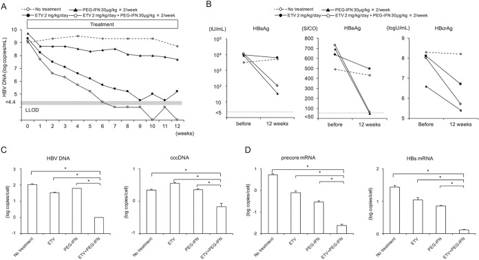
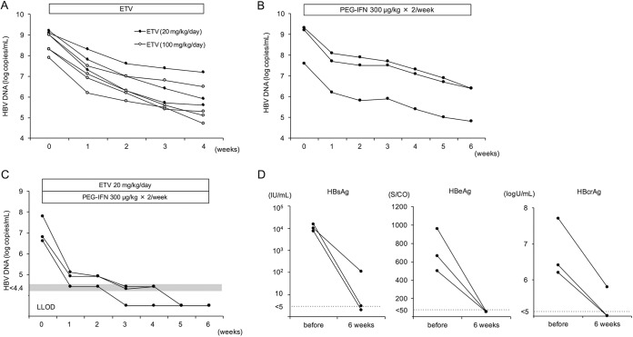
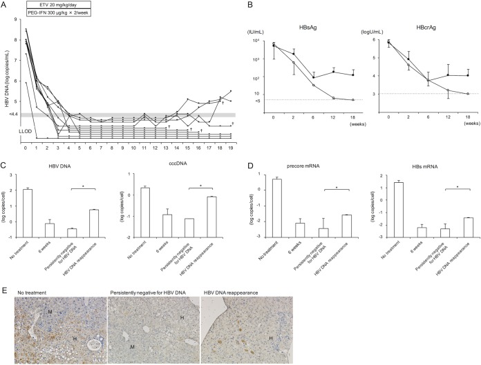
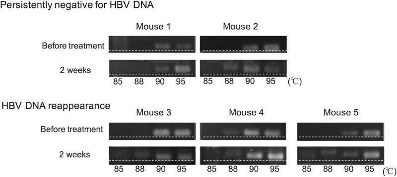
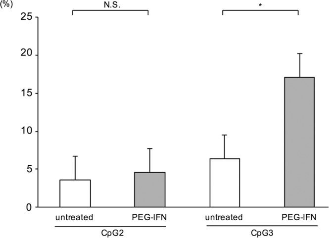
Nucleot(s)ide analogues and peginterferon (PEG-IFN) treatment are the only approved therapies for chronic hepatitis B virus (HBV) infection. However, complete eradication of the virus, as indicated by persistent loss of hepatitis B surface antigen (HBsAg), is rare among treated patients. This is due to long-term persistence of the HBV genome in infected hepatocytes in the form of covalently closed circular DNA (cccDNA). In this study, we investigated whether administration of a large dose of a nucleoside analogue in combination with PEG-IFN can achieve long-term loss of HBsAg in human hepatocyte chimeric mice. Mice were treated with a high dose of entecavir and/or PEG-IFN for 6 weeks. High-dose combination therapy with both drugs resulted in persistently negative HBV DNA in serum. Although small amounts of HBV DNA and cccDNA (0.1 and 0.01 copy/cell, respectively) remained in the mouse livers, some of the mice remained persistently negative for serum HBV DNA at 13 weeks after cessation of the therapy. Serum HBsAg and hepatitis B core-related antigen (HBcrAg) continued to decrease and eventually became negative at 12 weeks after cessation of the therapy. Analysis of the HBV genome in treated mice showed accumulation of G-to-A hypermutation and CpG III island methylation. Persistent loss of serum HBV DNA and loss of HBV markers by high-dose entecavir and PEG-IFN combination treatment in chimeric mice suggests that control of HBV can be achieved even in the absence of a cellular immune response.

Keywords: DNA methylation; cccDNA; hepatitis B virus; human hepatocyte chimeric mouse; hypermutation; methylation.


Figures

FIG 1
FIG 1
Effect of antivirals on HBV-infected mice. Four mice were treated with or without (no treatment) 2 mg/kg of entecavir (ETV) alone daily or 30 μg/kg of PEG-IFN alone or entecavir plus PEG-IFN twice weekly for 12 weeks. (A) Time course of serum HBV DNA titer in each mouse. (B) Serum HBsAg, HBeAg, and HBcrAg levels in each mouse at baseline and after 12 weeks of treatment. (C) Intrahepatic HBV DNA and HBV cccDNA levels in each mouse. An HBV-infected mouse without treatment (no treatment) was also analyzed. Data are presented as the mean ± standard deviation (SD) for three samples of liver tissue from each mouse. *, P < 0.05. (D) Intrahepatic precore mRNA and HBs mRNA. HBcrAg, hepatitis B core-related antigen. LLOD, lower limit of detection.
FIG 2
FIG 2
Changes in HBV markers in mice treated with high-dose antivirals. (A to B) HBV-infected mice were treated with either 20 mg/kg or 100 mg/kg of entecavir (ETV) daily for 4 weeks (A) or 300 μg/kg of PEG-IFN-α-2a twice weekly for 6 weeks (B). (C and D) Three mice were treated with 20 mg/kg of entecavir daily and 300 μg/kg of PEG-IFN twice weekly for 6 weeks. (C) The time course of serum HBV DNA titer in each mouse. (D) Serum HBsAg, HBeAg, and HBcrAg levels in each mouse at baseline and after 6 weeks of treatment in three mice.
FIG 3
FIG 3
Effect of high-dose antivirals on HBV-infected mice. Ten mice were treated with 20 mg/kg of entecavir daily and 300 μg/kg of PEG-IFN twice weekly for 6 weeks. (A) Time course of serum HBV DNA titer in each mouse. Mice in which serum HBV DNA reappeared after cessation of the treatment are shown with closed circles, and mice that remained persistently negative for serum HBV DNA after cessation of the treatment are shown with open circles. †, mice were lost at that time point, and therefore subsequent data could not be collected. Five mice were lost during examination, and the remaining five mice were sacrificed at 19 weeks. (B) Changes in serum HBsAg and HBcrAg levels. Mice in which serum HBV DNA reappeared after cessation of the treatment (closed circles) (n = 3) and mice that remained persistently negative for serum HBV DNA after cessation of the treatment (open circles) (n = 7) are shown. Data are presented as the mean ± SD. (C and D) Intrahepatic HBV DNA and HBV cccDNA (C) and precore mRNA and HBs mRNA (D) at 19 weeks in mice with serum HBV DNA reappearance after cessation of the treatment (HBV DNA reappearance; two samples of liver tissue in 2 mice) and mice persistently negative for serum HBV DNA after cessation of the treatment (persistently negative for HBV DNA; n = 3). HBV DNA, cccDNA, precore mRNA, and HBs mRNA levels were also measured in HBV-infected mice that did not receive treatment (no treatment; n = 6) or that received 6 weeks of combination treatment (6 weeks; n = 3). Data are presented as the mean ± SD. *, P < 0.05. (E) Histological analysis of liver samples. Liver samples were stained with anti-hepatitis B core (HBc) antibody. Labeled regions indicate human (H) and mouse (M) hepatocytes (original magnification, ×200). Similar histological patterns were observed in other mice in each group.
FIG 4
FIG 4
HBV hypermutation following treatment with PEG-IFN and entecavir. HBV DNA was amplified by 3D-PCR, and hypermutated genomes were detected by HA-yellow agarose gel electrophoresis in five mice that were positive for serum HBV DNA after 2 weeks of treatment with PEG-IFN plus entecavir. Serum HBV DNA remained persistently negative in two mice (persistently negative for HBV DNA; mice 1 and 2) and reappeared after cessation of the treatment in three mice (HBV DNA reappearance; mice 3, 4, and 5). More heavily hypermutated HBV DNA was observed at 88°C. The dashed lines were added to help visualize the retardation of AT-rich DNA in the HA-yellow agarose gel.
FIG 5
FIG 5
CpG methylation of the HBV genome following interferon treatment. Mice were treated with or without 300 μg/kg of PEG-IFN twice weekly. After 6 weeks of treatment, DNA was extracted from mouse livers, and the proportions of methylated sites in CpG II and CpG III islands were analyzed. Data are presented as the mean ± SD for three mice. *, P < 0.05; N.S., not significant.

Similar articles

Cited by

References

    1. Trépo C, Chan HLY, Lok A. 2014. Hepatitis B virus infection. Lancet 384:2053–2063. doi:10.1016/S0140-6736(14)60220-8. - DOI - PubMed
    1. Nassal M. 2015. HBV cccDNA: viral persistence reservoir and key obstacle for a cure of chronic hepatitis B. Gut 64:1972–1984. doi:10.1136/gutjnl-2015-309809. - DOI - PubMed
    1. Marcellin P, Gane E, Buti M, Afdhal N, Sievert W, Jacobson IM, Washington MK, Germanidis G, Flaherty JF, Schall RA, Bornstein JD, Kitrinos KM, Subramanian GM, McHutchison JG, Heathcote EJ. 2013. Regression of cirrhosis during treatment with tenofovir disoproxil fumarate for chronic hepatitis B: a 5-year open-label follow-up study. Lancet 381:468–475. doi:10.1016/S0140-6736(12)61425-1. - DOI - PubMed
    1. Chang TT, Liaw YF, Wu SS, Schiff E, Han KH, Lai CL, Safadi R, Lee SS, Halota W, Goodman Z, Chi YC, Zhang H, Hindes R, Iloeje U, Beebe S, Kreter B. 2010. Long-term entecavir therapy results in the reversal of fibrosis/cirrhosis and continued histological improvement in patients with chronic hepatitis B. Hepatology 52:886–893. doi:10.1002/hep.23785. - DOI - PubMed
    1. Dienstag JL, Goldin RD, Heathcote EJ, Hann HWL, Woessner M, Stephenson SL, Gardner S, Gray DF, Schiff ER. 2003. Histological outcome during long-term lamivudine therapy. Gastroenterology 124:105–117. doi:10.1053/gast.2003.50013. - DOI - PubMed

MeSH terms

LinkOut - more resources