. 2017 Jul 25;114(30):E6072-E6078.
doi: 10.1073/pnas.1702295114. Epub 2017 Jul 10.

Functional characterization of human pluripotent stem cell-derived arterial endothelial cells

Affiliations

Functional characterization of human pluripotent stem cell-derived arterial endothelial cells

Jue Zhang et al. Proc Natl Acad Sci U S A. .

Abstract

Here, we report the derivation of arterial endothelial cells from human pluripotent stem cells that exhibit arterial-specific functions in vitro and in vivo. We combine single-cell RNA sequencing of embryonic mouse endothelial cells with an EFNB2-tdTomato/EPHB4-EGFP dual reporter human embryonic stem cell line to identify factors that regulate arterial endothelial cell specification. The resulting xeno-free protocol produces cells with gene expression profiles, oxygen consumption rates, nitric oxide production levels, shear stress responses, and TNFα-induced leukocyte adhesion rates characteristic of arterial endothelial cells. Arterial endothelial cells were robustly generated from multiple human embryonic and induced pluripotent stem cell lines and have potential applications for both disease modeling and regenerative medicine.

Keywords: arterial endothelial cells; arterial-specific functions; human pluripotent stem cell differentiation; myocardial infarction; single-cell RNA-seq.


Conflict of interest statement

Conflict of interest statement: W.L.M. is a founder and stockholder for Stem Pharm Inc. and Tissue Regeneration Systems Inc.

Figures

Fig. 1.
Fig. 1.
Identification of candidate factors critical to arteriovenous specification. (A and C) Flow cytometric analysis of CD31, CD144, EFNB2-tdTomato (EFNB2-Tom), and EPHB4-EGFP expression. The gate setting is based on undifferentiated embryonic stem cells for all of the flow cytometric data in this study. EFNB2-tdTomato/EPHB4-EGFP dual reporter cells were first differentiated to mesoderm cells using E8BAC media [E8 media (45) supplemented with 5 ng/mL BMP4, 25 ng/mL Activin A, and 1 μM CHIR99021]. E5 (E8 media minus FGF2, TGFβ1, and insulin) media supplemented with 100 ng/mL FGF2, 50 ng/mL VEGFA, and 50 ng/mL BMP4 was used to induce mesoderm cells to differentiate into endothelial cells from day 2 to day 6. Insulin (20 μg/mL) was added to or removed from the media from day 2 to day 6 as indicated. (B and D) Statistics of CD31+CD144+ and EFNB2-tdTomatohigh/EPHB4-EGFPlow cells. Data are represented as mean ± SD. Student’s t test; *P < 0.05, n = 3. (E) Flow cytometric analysis of EFNB2-tdTomato and EPHB4-EGFP expression on CD31 and CD144 gated endothelial cells. E5 media supplemented with 50 ng/mL VEGFA and 10 μM SB431542 was used to induce mesoderm cells differentiated into endothelial cells from day 2 to day 6. BMP4 (50 ng/mL) was added as indicated. Experiments were repeated three times. (F and H) Flow cytometric analysis of CD31+CD144+ and EFNB2-tdTomatohigh/EPHB4-EGFPlow cells. Embryonic stem cells were first differentiated into mesoderm cells as mentioned above. E5 media supplemented with 50 ng/mL VEGFA and 10 μM SB431542 was used as the base media to induce mesoderm cells to differentiate into endothelial cells from day 2 to day 6. Other factors were added to the base media as indicated. (G and I) Statistics of CD31+CD144+ and EFNB2-tdTomatohigh/EPHB4-EGFPlow cells. Data are represented as mean ± SD. Student’s t test; *P < 0.05; n = 3. The following were used: 5 μM L690, 5 μg/mL LDL, and 100 ng/mL PDGF-BB.
Fig. 2.
Fig. 2.
Combinatorial effects of the key factors. (A–D) Flow cytometric analysis of CD31+CD144+, EFNB2-tdTomatohigh/EPHB4-EGFPlow, CD144+CXCR4+, and CD144+DLL4+ cells. Five factor media (E5 media supplemented with 100 ng/mL FGF2, 50 ng/mL VEGFA, 10 μM SB431542, 5 μM RESV, and 5 μM L690) was used as the base media. As indicated, single factors were either withdrawn from or added into the five factor media. Insulin, 20 μg/mL. Statistical data of CD31+CD144+, EFNB2-tdTomatohigh/EPHB4-EGFPlow, CD144+CXCR4+, and CD144+DLL4+ cells are represented as mean ± SD. Student’s t test; *P < 0.05; n = 3. (E) Statistics of AEC number generated from 1.0 × 10⁶ starting embryonic stem (ES) cells or one 10-cm dish with 8.3 × 10⁶ starting ES cells after 6 d of differentiation (five factor media). n = 3. (F) A schematic of five factor protocol. EFNB2-Tom, EFNB2-tdTomato.
Fig. 3.
Fig. 3.
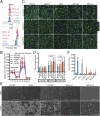
Characterization of AECs. (A and B) Flow cytometric analysis of the expression of EFNB2-tdTomato, EPHB4-EGFP, DLL4, and CXCR4. The experiment was performed three times; one of the typical results is shown. AECs were derived using the five factor protocol. VECs were derived using E6 media (E8 media minus FGF2 and TGF-β1, containing insulin) supplemented with 50 ng/mL VEGFA and 50 ng/mL BMP4 to differentiate mesoderm cells into endothelial cells from day 2–6. (C) qRT-PCR of arterial and venous markers. AECs and VECs were purified by flow cytometry. Data are represented as mean ± SD. Student’s t test; *P < 0.05; n = 3. (D) Acetylated LDL (Ac-LDL) uptake. (Scale bar, 100 μm.) AECs (passage 2) derived from wild-type H1 cells were used. The purity was ∼93% after being passaged, so cells used in C–H were not purified. (E) Matrigel encapsulation assay. AECs (passage 3) derived from the reporter cell line were used. (Scale bar, 100 μm.) (F) Immunostaining of NICD1 (activated). (Scale bar, 50 μm.) AECs derived from wild-type H1 cells were used for F–H. (G) Vascular formation in fibrin gel. (Scale bar, 100 μm.) (H) Lumen formation of endothelial cell and pericyte cocultured in fibrin gel. To visualize the lumen, cells were stained with CMFDA (green). Y-z and x-z projection was shown. (Scale bar, 100 μm.) (I) Endothelial cells formed functional vessels in vivo. Wild-type H1-derived AECs (passage 2, purified by CD144 microbeads) were mixed with Matrigel and injected into SCID mice. After 4 wk, rhodamine-dextran was retro-orbital injected to highlight perfused vessels. (Scale bar, 100 μm.) CD31, the antibody recognizes both human and mouse CD31. hCD144, anti-human CD144 antibody.
Fig. 4.
Fig. 4.
Arterial-specific functional characterization of endothelial cells. (A) NO production was revealed by the intensity of DAF-FM. AECs were derived from wild-type H1 cells using the five factor protocol and used for experiments at passage 2 or 4 as indicated. VECs were derived using E6 media (E8 media minus FGF2 and TGF-β1) supplemented with 50 ng/mL VEGFA and 50 ng/mL BMP4 to induce mesoderm cells differentiated into endothelial cells from day 2–6. CD31+ CD144+ cells were purified for the experiments. DAF-FM is nonfluorescent until it reacts with NO to form a fluorescent benzotriazole. The fluorescent intensity was measure by flow cytometry. The experiment was performed three times; one of the typical results is shown. (B) Oxygen consumption rate was measured on XF24 analyzers (Seahorse Bioscience). Oligomycin was used to abolish the oxygen consumption. FCCP was used to uncouple the electron transport chain from the oxidative phosphorylation, thus measuring the maximal respiration capacity. Antimycin A and Rotenone were applied simultaneously to completely block the electron transport chain. Student’s t test; *P < 0.05; n = 4. The P value was calculated by comparing to HUVECs. (C) Shear stress response was performed on ibidi Pump System (Perfusion Set RED, μ-Slide VI 0.4). (D) Statistical data of shear stress response. Ratio of cell length to width was used to demonstrate the elongation of cells in response to shear stress. For each cell type, 100 cells were measured to do the statistics. Data are represented as mean ± SD. Student’s t test; *P < 0.05; n = 100 cells from three independent experiments. (E) Leukocyte (round cells) adhesion assay. (Scale bar, 200 μm.) (F) Statistics of leukocyte adhesion assay. Leukocyte number was counted for each image. Data are represented as mean ± SD. Student’s t test; *P < 0.05; n = 3 images from three independent experiments. The P value was calculated by comparing to HUVECs with TNFα treatment. –, nontreated; +, TNFα treated.
Fig. 5.
Fig. 5.
AECs improve cardiovascular function. (A) Survival rate of the mice. AECs group: n = 12, 2.5 × 10⁶ AECs were injected per mouse. VECs group: n = 8, 2.5 × 10 (30) VECs were injected per mouse. PBS group: n = 12. (B) Kaplan–Meier survival curve. *P < 0.05. The P value between the AECs and PBS group or the VECs and PBS group was calculated by Mantel–Cox test. AECs group, n = 12. VECs group, n = 8. PBS group, n = 12. (C) Immunostaining. AECs and VECs were stained with human-specific CD31 antibody. Alexa Fluor 647 Hydrazide (1 μg/mL) was used to label the elastin produced by arteries (46). hCD31, anti-human CD31 antibody. (Scale bar, 50 μm.)

References

    1. Go AS, et al. American Heart Association Statistics Committee and Stroke Statistics Subcommittee Heart disease and stroke statistics–2014 update: A report from the American Heart Association. Circulation. 2014;129:e28–e292. - PMC - PubMed
    1. Epstein AJ, Polsky D, Yang F, Yang L, Groeneveld PW. Coronary revascularization trends in the United States, 2001-2008. JAMA. 2011;305:1769–1776. - PMC - PubMed
    1. Goodney PP, Beck AW, Nagle J, Welch HG, Zwolak RM. National trends in lower extremity bypass surgery, endovascular interventions, and major amputations. J Vasc Surg. 2009;50:54–60. - PubMed
    1. Campbell GR, Campbell JH. Development of tissue engineered vascular grafts. Curr Pharm Biotechnol. 2007;8:43–50. - PubMed
    1. Aranguren XL, et al. Unraveling a novel transcription factor code determining the human arterial-specific endothelial cell signature. Blood. 2013;122:3982–3992. - PubMed

Publication types