. 2018 Mar;23(3):691-700.
doi: 10.1038/mp.2017.137. Epub 2017 Jul 11.

GluD1, linked to schizophrenia, controls the burst firing of dopamine neurons

Affiliations

GluD1, linked to schizophrenia, controls the burst firing of dopamine neurons

N Benamer et al. Mol Psychiatry. 2018 Mar.

Abstract

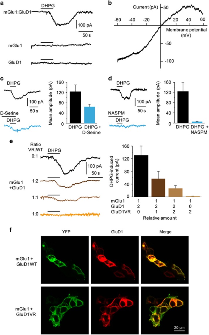
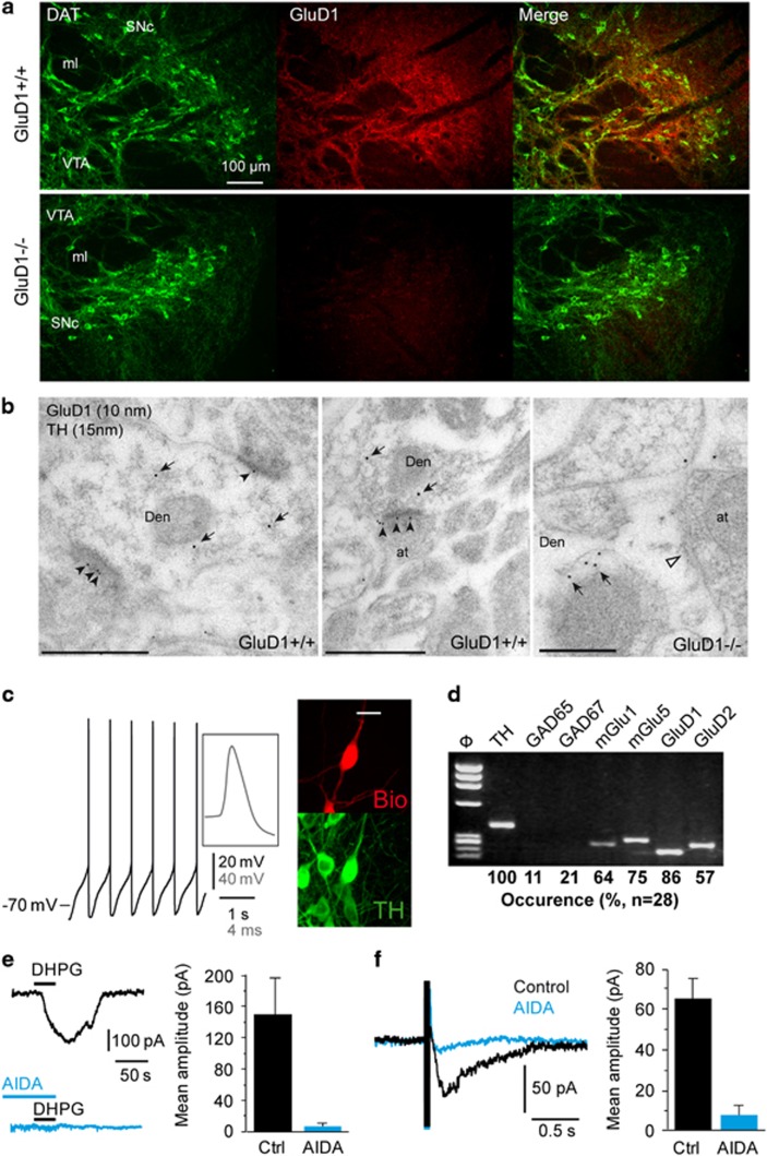
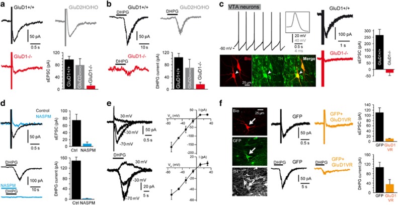
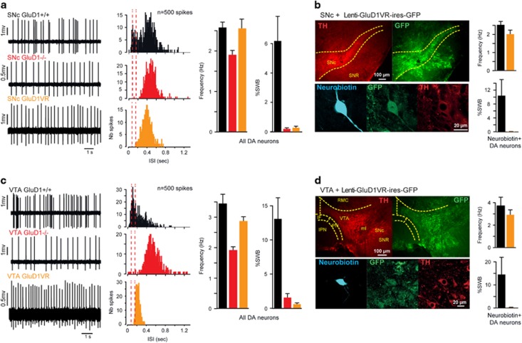
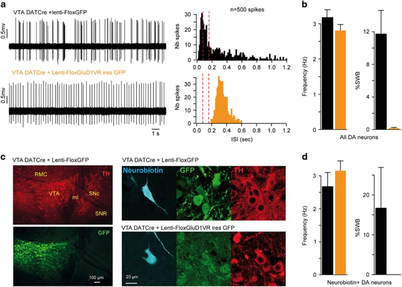
Human mutations of the GRID1 gene encoding the orphan delta1 glutamate receptor-channel (GluD1) are associated with schizophrenia but the explicit role of GluD1 in brain circuits is unknown. Based on the known function of its paralog GluD2 in cerebellum, we searched for a role of GluD1 in slow glutamatergic transmission mediated by metabotropic receptor mGlu1 in midbrain dopamine neurons, whose dysfunction is a hallmark of schizophrenia. We found that an mGlu1 agonist elicits a slow depolarizing current in HEK cells co-expressing mGlu1 and GluD1, but not in cells expressing mGlu1 or GluD1 alone. This current is abolished by additional co-expression of a dominant-negative GluD1 dead pore mutant. We then characterized mGlu1-dependent currents in dopamine neurons from midbrain slices. Both the agonist-evoked and the slow postsynaptic currents are abolished by expression of the dominant-negative GluD1 mutant, pointing to the involvement of native GluD1 channels in these currents. Likewise, both mGlu1-dependent currents are suppressed in GRID1 knockout mice, which reportedly display endophenotypes relevant for schizophrenia. It is known that mGlu1 activation triggers the transition from tonic to burst firing of dopamine neurons, which signals salient stimuli and encodes reward prediction. In vivo recordings of dopamine neurons showed that their spontaneous burst firing is abolished in GRID1 knockout mice or upon targeted expression of the dominant-negative GluD1 mutant in wild-type mice. Our results de-orphanize GluD1, unravel its key role in slow glutamatergic transmission and provide insights into how GRID1 gene alterations can lead to dopaminergic dysfunctions in schizophrenia.


Conflict of interest statement

The authors declare no conflict of interest.

Figures

Figure 1
Figure 1
Activation of mGlu1 triggers the opening of GluD1 channels in HEK cells. (a) Bath application of DHPG (100 μM) induced a slow inward current in cells co-expressing mGlu1 and GluD1, but not in cells expressing either mGlu1 or GluD1 alone. (b) Example of I/V curve of the DHPG-induced current exhibiting a reversal potential around 0 mV and inward rectification at positive potentials. (c,d) The DHPG-induced current was reduced by D-serine (10 mM) and almost abolished by NASPM (100 μM). (e) Co-expression of the dominant-negative GluD1VR dead pore mutant with mGlu1 and WT GluD1 dose-dependently reduced the DHPG-induced current. (f) Correct targeting of recombinant proteins at the plasma membrane. DHPG, 3,5-dihydroxyphenylglycine; NASPM, naphtyl-acetyl spermine; WT, wild type.
Figure 2
Figure 2
GluD1 is co-expressed with mGlu1/5 in DA neurons. (a) Fluorescence micrographs of the SNc and VTA showing diffuse GluD1 immunostaining of somata and processes of DAT-positive DA neurons. Immunostaining almost disappeared in GluD1−/− mice. Scale bar: 100 μm. (b) Electron micrographs of the SNc showing immunoparticles for GluD1 (arrowheads) and TH (arrows), as detected using a double-labeling post-embedding immunogold method. GluD1 immunoparticles were detected along the postsynaptic density of dendritic shafts (Den) of TH-labeled DA neurons, establishing asymmetrical synapses with axon terminal (at). No GluD1 immunoparticles were detected in GluD1−/− mice, as highlighted by open arrowhead pointing to postsynaptic density of a TH-labeled neuron. Scale bars: 0.5 μm. (c) Characterization of a DA neuron from the SNc typically exhibiting long duration action potentials and spontaneous pacemaker firing at low rate. This biocytin-filled neuron (Bio) was TH-immunopositive (scale bar: 20 μm). (d) RT-PCR analysis of a single DA neuron detected expression of TH, mGlu1, mGlu5, GluD1 and GluD2 upon agarose gel electrophoresis of PCR products (ΦX HaeIII molecular weight marker). Summary of single-cell RT-PCR data obtained in SNc DA neurons is given below the agarose gel picture. (e,f) The mGlu1/5 antagonist AIDA (150 μM) prevented the DHPG-induced current and the sEPSC triggered by local electrical stimulation in DA neurons. AIDA, 1-aminoindan-1,5-dicarboxylic acid; DA, dopamine; RT-PCR, real-time PCR; sEPCS, slow excitatory postsynaptic current; SN, substantia nigra; SNc, SN pars compacta; VTA, ventral tegmental area.
Figure 3
Figure 3
GluD1 channels mediate mGlu1/5-dependent currents in DA neurons. (a,b) The sEPSC and the IDHPG of SNc DA neurons were strongly reduced in GluD1−/− mice, but were not significantly altered in mice bearing a deletion of the GluD2 gene (GluD2HO/HO). (c) In the VTA, GluD1 deletion also hampered the sEPSC (right panels) of DA neurons identified from their long duration action potentials, spontaneous pacemaker firing at low rate and TH immunoreactivity (left panels). Traces above fluorescence pictures stem from recording of the biocytin-filled cell indicated by an arrow. (d) Application of the GluD channel blocker NASPM (100 μM) almost abolished the sEPSC and the IDHPG in DA neurons of the SNc. (e) I/V curves of the sEPSC and the IDHPG recorded at different preset potentials in DA neurons of the SNc, as exemplified by superimposed traces in left panels. (f) Fluorescence pictures show histological analysis of a GFP-positive DA neuron recorded 20 h after injection of a Sindbis virus co-expressing GluD1VR and GFP in the SNc of a WT mouse and labeled with biocytin (Bio). Note the sparse transduction achieved using sindbis viral transfer in the SNc. The sEPSC and the IDHPG were largely reduced in SNc DA neurons co-expressing GluD1VR and GFP compared to DA neurons transduced with a control Sindbis virus expressing GFP alone. DA, dopamine; NASPM, naphtyl-acetyl spermine; sEPCS, slow excitatory postsynaptic current; SNc, SN pars compacta; SNR, substantia nigra pars reticulata; VTA, ventral tegmental area; WT, wild type.
Figure 4
Figure 4
GluD1 controls the burst firing of DA neurons in vivo. (a,c) Examples of spontaneous spike firing patterns (left panels) and corresponding histograms of ISI (middle panels, red dotted lines indicate the limits of burst onset <80 ms and termination >160 ms) obtained from DA neurons recorded in GluD1+/+ and GluD1−/− mice and in WT mice injected with a lentivirus co-expressing GluD1VR and GFP in the SNc or VTA. Right panels show the mean spike frequency and %SWB of recorded DA neurons. Note that burst firing of DA neurons is virtually abolished by global (GluD1−/−) or local (GluD1VR) genetic disruption of GluD1. (b,d) Left panels: Histological analysis of virally transduced DA neurons recorded 4–6 weeks after injection of lentivirus LV-PGK-GluD1V617R-ires-GFP and filled with neurobiotin. DA, dopamine; IF, interfascicular nucleus; IPN, interpeduncular nucleus; ISI, interspike intervals; ml, medial lemniscus; PN, paranigral nucleus; RMC, red nucleus; SNc, SN pars compacta; SNR, substantia nigra pars reticulata; VTA, ventral tegmental area; WT, wild type; %SWB, percentage of spikes within burst. Right panels: same analyses as in (a,c) restricted to neurobiotin+/TH+ (GluD1+/+) and neurobiotin+/GFP+/TH+ (GluD1VR) recorded neurons.
Figure 5
Figure 5
Selective expression of GluD1VR mutant in DA neurons suppresses burst firing. (a) Examples of spontaneous spike firing patterns (left panels) and corresponding histograms of ISI (middle panels) obtained from VTA DA neurons recorded in DAT-Cre mice after injection of a Cre-dependent lentivirus expressing either GFP, or GluD1VR and GFP, in the VTA. (b) Mean spike frequency and percentage of spikes within burst of recorded DA neurons. Note that selective expression of GluD1VR in DA neurons abolishes burst firing without significant effect on firing frequency. (c) Left: Whole field fluorescence micrographs showing selective expression of GFP in VTA TH+ neurons after viral transduction. Right: histological analysis of virally transduced DA neurons recorded 6 weeks after injection of indicated lentivirus and filled with neurobiotin. The oblique neurobiotin-labeled dendrite in lower left picture belongs to another GFP+/TH+ recorded neuron whose soma is out of field. (d) Same analyses as in (b) restricted to neurobiotin+/GFP+/TH+ recorded neurons. DA, dopamine; ISI, interspike intervals; VTA, ventral tegmental area;.

References

    1. Fallin MD, Lasseter VK, Avramopoulos D, Nicodemus KK, Wolyniec PS, McGrath JA et al. Bipolar I disorder and schizophrenia: a 440-single-nucleotide polymorphism screen of 64 candidate genes among Ashkenazi Jewish case-parent trios. Am J Hum Genet 2005; 77: 918–936. - PMC - PubMed
    1. Greenwood TA, Lazzeroni LC, Murray SS, Cadenhead KS, Calkins ME, Dobie DJ et al. Analysis of 94 candidate genes and 12 endophenotypes for schizophrenia from the Consortium on the Genetics of Schizophrenia. Am J Psychiatry 2011; 168: 930–946. - PMC - PubMed
    1. Guo SZ, Huang K, Shi YY, Tang W, Zhou J, Feng GY et al. A case-control association study between the GRID1 gene and schizophrenia in the Chinese Northern Han population. Schizophr Res 2007; 93: 385–390. - PubMed
    1. Treutlein J, Muhleisen TW, Frank J, Mattheisen M, Herms S, Ludwig KU et al. Dissection of phenotype reveals possible association between schizophrenia and Glutamate Receptor Delta 1 (GRID1) gene promoter. Schizophr Res 2009; 111: 123–130. - PubMed
    1. Gao J, Maison SF, Wu X, Hirose K, Jones SM, Bayazitov I et al. Orphan glutamate receptor delta1 subunit required for high-frequency hearing. Mol Cell Biol 2007; 27: 4500–4512. - PMC - PubMed

Publication types