. 2017 Jul 1;22(7):76006.
doi: 10.1117/1.JBO.22.7.076006.

Handheld optical coherence tomography-reflectance confocal microscopy probe for detection of basal cell carcinoma and delineation of margins

Affiliations

Handheld optical coherence tomography-reflectance confocal microscopy probe for detection of basal cell carcinoma and delineation of margins

Nicusor Iftimia et al. J Biomed Opt. .

Abstract

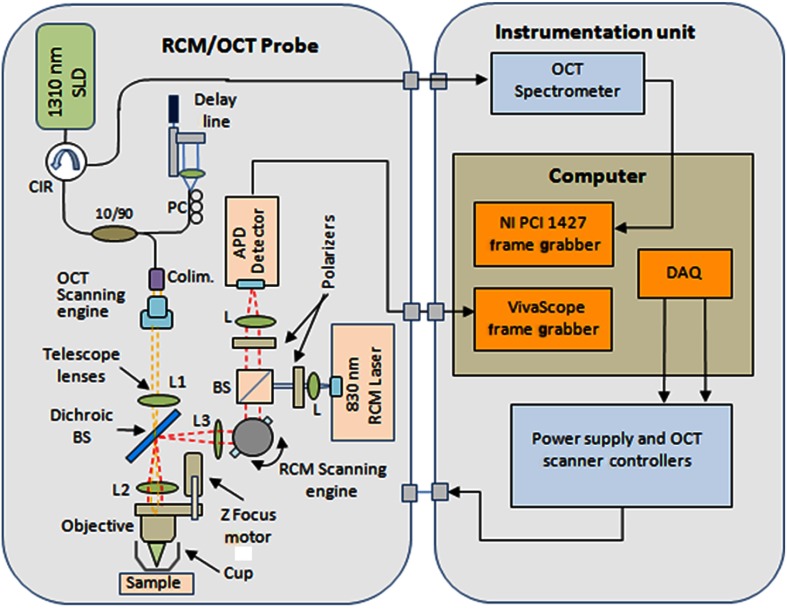
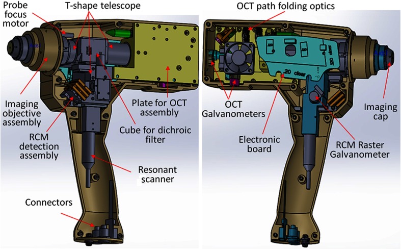
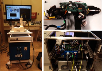
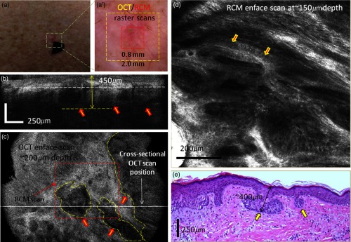
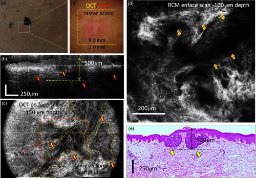
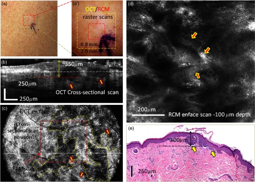
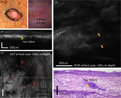
We present a hand-held implementation and preliminary evaluation of a combined optical coherence tomography (OCT) and reflectance confocal microscopy (RCM) probe for detecting and delineating the margins of basal cell carcinomas (BCCs) in human skin <italic<in vivo</italic<. A standard OCT approach (spectrometer-based) with a central wavelength of 1310 nm and 0.11 numerical aperture (NA) was combined with a standard RCM approach (830-nm wavelength and 0.9 NA) into a common path hand-held probe. Cross-sectional OCT images and enface RCM images are simultaneously displayed, allowing for three-dimensional microscopic assessment of tumor morphology in real time. Depending on the subtype and depth of the BCC tumor and surrounding skin conditions, OCT and RCM imaging are able to complement each other, the strengths of each helping overcome the limitations of the other. Four representative cases are summarized, out of the 15 investigated in a preliminary pilot study, demonstrating how OCT and RCM imaging may be synergistically combined to more accurately detect BCCs and more completely delineate margins. Our preliminary results highlight the potential benefits of combining the two technologies within a single probe to potentially guide diagnosis as well as treatment of BCCs.


Figures

Fig. 1
Fig. 1
Simplified schematic of the RCM/OCT instrument.
Fig. 2
Fig. 2
Solid Work (CAD) design of the handheld OCT/RCM probe.
Fig. 3
Fig. 3
Photographs of the RCM/OCT imaging instrument: (a) general view of the instrumentation unit and hand-held probe; (b) probe detail; and (c) instrumentation unit detail.
Fig. 4
Fig. 4
(a) Clinical image showing an erythematous macule on the back. (a′) Dermoscopy image showing shiny white lines and serpentine vessels. (b) Cross-sectional and (c) enface OCT images showing multiple hypoechoic areas (see arrows), suggestive of BCC. (d) Reflectance confocal microscopy showing cord-like structures with peripheral palisading (arrows) admixed with a fibrotic stroma, suggestive of BCC. (e) Histology image of the lesion (confirming the diagnosis of superficial BCC), showing multiple small tumor nests originating from the epidermis (hematoxylin and eosin, 4× magnification).
Fig. 5
Fig. 5
(a) Clinical image showing an erythematous macule on the right shoulder. (a′) Dermoscopy image showing shiny white lines and serpentine vessels, suggestive of superficial BCC. (b) Cross-sectional and (c) enface OCT images showing multiple hypoechoic areas, suggestive of BCC. (d) Reflectance confocal microscopy showing cord-like structures with palisading (arrows) surrounded by reticulated collagen and inflammatory cells. (e) Histology showing multiple tumor nests with different sizes originating from the epidermis (arrows) (hematoxylin and eosin, 4× magnification).
Fig. 6
Fig. 6
(a) Clinical image showing a subtle whitish macule on the left arm. (a′) Dermoscopy image showing shiny white lines and few serpentine vessels. (b) Cross-sectional and (c) enface OCT images show multiple hypoechoic areas suggestive of BCC. (d) Reflectance confocal microscopy image showing cord-like structures without clear palisading (arrows). (e) Histology image of the lesion revealing multiple small tumor nests originating from the epidermis confirming the diagnosis of superficial BCC (hematoxylin and eosin, 4× magnification).
Fig. 7
Fig. 7
(a) Clinical image showing a subtle whitish macule on a female’s leg. (a′) Dermoscopy image showing subtle white lines, rainbow pattern, and a few vessels. (b) Cross-sectional and (c) enface OCT images showed hypoechoic areas corresponding to branched blood vessels, and a hypoechoic nodular area surrounded by a hyper-reflective halo corresponding to a hair follicle. (d) Reflectance confocal microscopy image showing bundled collagen (arrows) but no cordlike structures suggestive of BCC. (e) Histology image of the lesion revealing a hair follicle (see arrowhead highlighting the hair) and bundled collagen corresponding to fibrosis secondary to trauma (hematoxylin and eosin, 4× magnification).

Similar articles

Cited by

References

    1. Kadouch D. J., et al. , “In vivo confocal microscopy of basal cell carcinoma: a systematic review of diagnostic accuracy,” J. Eur. Acad. Dermatol. Venereol. 29(10), 1890–1897 (2015).JEAVEQ10.1111/jdv.13224 - DOI - PubMed
    1. Xiong Y. D., et al. , “A meta-analysis of reflectance confocal microscopy for the diagnosis of malignant skin tumours,” J. Eur. Acad. Dermatol. Venereol. 30, 1295–1302 (2016).10.1111/jdv.2016.30.issue-8 - DOI - PubMed
    1. Cheng H. M., Guitera P., “Systematic review of optical coherence tomography usage in the diagnosis and management of basal cell carcinoma,” Br. J. Dermatol. 173(6), 1371–1380 (2015).10.1111/bjd.14042 - DOI - PubMed
    1. Cheng H. M., et al. , “Accuracy of optical coherence tomography for the diagnosis of superficial basal cell carcinoma—a prospective, consecutive, cohort study of 168 cases,” Br. J. Dermatol. 175(6), 1290–1300 (2016).10.1111/bjd.14714 - DOI - PubMed
    1. Huang D., et al. , “Optical coherence tomography,” Science 254, 1178–1181 (1991).SCIEAS10.1126/science.1957169 - DOI - PMC - PubMed