. 2017 Jul 11;12(1):54.
doi: 10.1186/s13024-017-0193-9.

A proteomic analysis of LRRK2 binding partners reveals interactions with multiple signaling components of the WNT/PCP pathway

Affiliations

A proteomic analysis of LRRK2 binding partners reveals interactions with multiple signaling components of the WNT/PCP pathway

Alena Salašová et al. Mol Neurodegener. .

Abstract

Background: Autosomal-dominant mutations in the Park8 gene encoding Leucine-rich repeat kinase 2 (LRRK2) have been identified to cause up to 40% of the genetic forms of Parkinson's disease. However, the function and molecular pathways regulated by LRRK2 are largely unknown. It has been shown that LRRK2 serves as a scaffold during activation of WNT/β-catenin signaling via its interaction with the β-catenin destruction complex, DVL1-3 and LRP6. In this study, we examine whether LRRK2 also interacts with signaling components of the WNT/Planar Cell Polarity (WNT/PCP) pathway, which controls the maturation of substantia nigra dopaminergic neurons, the main cell type lost in Parkinson's disease patients.

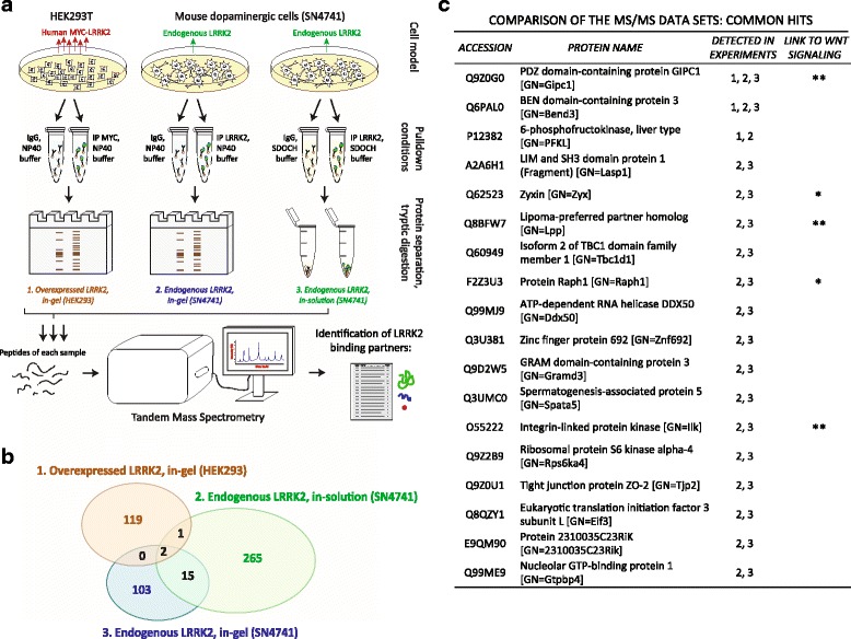
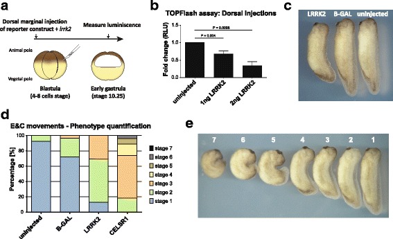
Methods: Co-immunoprecipitation and tandem mass spectrometry was performed in a mouse substantia nigra cell line (SN4741) and human HEK293T cell line in order to identify novel LRRK2 binding partners. Inhibition of the WNT/β-catenin reporter, TOPFlash, was used as a read-out of WNT/PCP pathway activation. The capacity of LRRK2 to regulate WNT/PCP signaling in vivo was tested in Xenopus laevis' early development.

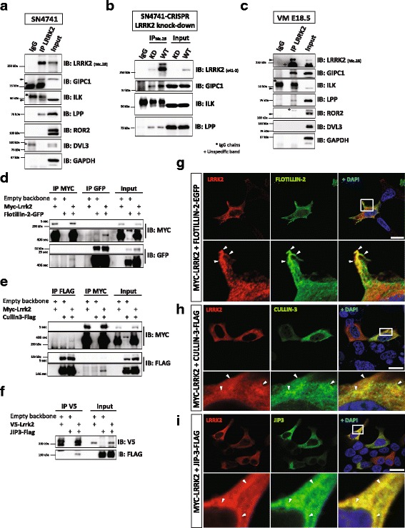
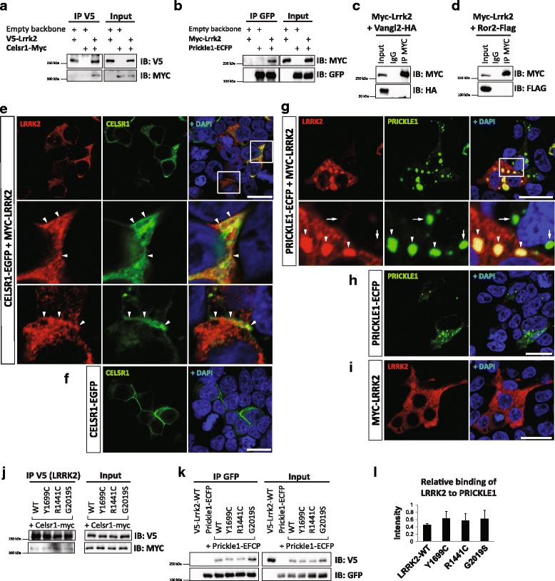
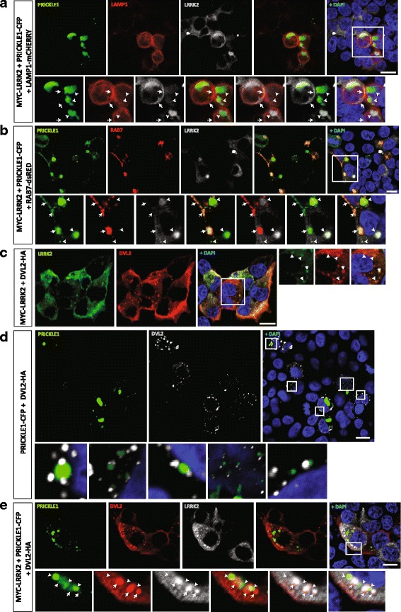
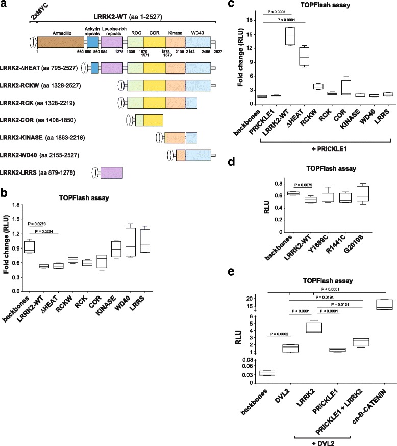
Results: Our proteomic analysis identified that LRRK2 interacts with proteins involved in WNT/PCP signaling such as the PDZ domain-containing protein GIPC1 and Integrin-linked kinase (ILK) in dopaminergic cells in vitro and in the mouse ventral midbrain in vivo. Moreover, co-immunoprecipitation analysis revealed that LRRK2 binds to two core components of the WNT/PCP signaling pathway, PRICKLE1 and CELSR1, as well as to FLOTILLIN-2 and CULLIN-3, which regulate WNT secretion and inhibit WNT/β-catenin signaling, respectively. We also found that PRICKLE1 and LRRK2 localize in signalosomes and act as dual regulators of WNT/PCP and β-catenin signaling. Accordingly, analysis of the function of LRRK2 in vivo, in X. laevis revelaed that LRKK2 not only inhibits WNT/β-catenin pathway, but induces a classical WNT/PCP phenotype in vivo.

Conclusions: Our study shows for the first time that LRRK2 activates the WNT/PCP signaling pathway through its interaction to multiple WNT/PCP components. We suggest that LRRK2 regulates the balance between WNT/β-catenin and WNT/PCP signaling, depending on the binding partners. Since this balance is crucial for homeostasis of midbrain dopaminergic neurons, we hypothesize that its alteration may contribute to the pathophysiology of Parkinson's disease.

Keywords: CELSR1; DVL; Dopaminergic neurons; Endocytosis; Immunoprecipitation; PRICKLE1; Parkinson’s disease; Signalosomes; Substantia nigra; WNT/planar cell polarity.


Conflict of interest statement

Ethics approval

Mice were housed, bred and treated according to the guidelines of the European Communities Council (directive 86/609/EEC) and the local ethics committees (Stockholm’s Norra Djurförsöketiska Nämnd N158/15). Xenopus laevis were housed and treated following institutional guidelines for animal care and research protocols approved by the local ethical committee (Stockholm’s Norra Djurförsöketiska Nämnd, permit N241/14). The lentiviruses were produced and handled according to the guidlines of the local ethical committee (Arbetsmiljöverket, permit Dnr 2-5164/2016).

Consent for publication

Not applicable.

Competing interests

The authors declare that they have no conflict of interest.

Publisher’s Note

Springer Nature remains neutral with regard to jurisdictional claims in published maps and institutional affiliations.

Figures

Fig. 1
Fig. 1
Large-scale immunoprecipitation-coupled MS/MS screening of LRRK2 binding partners revealed proteins belonging to WNT/PCP signaling pathway. a Scheme of the experimental workflow. b Venn diagram showing results from 3 different MS/MS data sets. c A list of identified proteins which were detected at least in 2 experiments. Proteins that have been linked to WNT signaling are marked with a star, stronger evidence is denoted by with two stars
Fig. 2
Fig. 2
Verification of the MS results in vitro and in vivo in mouse ventral midbrain. (a-c) Western blot validation of LRRK2 binding partners using specific antibodies. ILK and GIPC1 but no other WNT/PCP signaling components interact with LRRK2 in SN4741 (a-b) and mouse ventral midbrain of E18.5 old embryos (c). b We knocked down LRRK2 using CRISPR/Cas9 methodology and generated clonal cell lines with either normal (WT) or decreased levels of LRRK2 (KD). SN4741-LRRK2-KD served as a negative control for our co-IP experiments. d-f Western blot analysis of co-IP of transiently overexpressed candidates shows that LRRK2 binds to FLOTILLIN-2 (d) and CULLIN-3 (e) in HEK293T. JIP3 (f), a previously published LRRK2 binding partner, served as positive control. g-i IF confirmed that LRRK2 co-localizes with FLOTILLIN-2 (g), CULLIN-3 (h), and JIP3 (i) in HEK293T cells. Nuclear staining by Dapi is in blue. N ≥ 3. Scale bars indicate 20 μm
Fig. 3
Fig. 3
LRRK2 interacts with CELSR1 and PRICKLE1, two key components of WNT/PCP signaling. a-d Western blotting analysis of co-IP of transiently overexpressed LRRK2 with WNT/PCP signaling components in HEK293T. LRRK2 physically interacts with CELSR1 (a) and PRICKLE1 (b), but neither with VANGL2 (c) nor ROR2 (d). e-i IF of proteins overexpressed in HEK293T. Under normal conditions, LRRK2 is evenly distributed in the cytoplasm (i). CELSR1 alone is usually polarized in cells (f), whereas PRICKLE1 tends to form puncta (h). e Once co-expressed, LRRK2 partially co-localizes with CELSR1 in the cytoplasmic membrane and in cell-cell contacts (arrowheads). LRRK2 robustly changes its localization when it is co-expressed with PRICKLE1, and together they form puncta structures in the cytoplasm (g). Arrowheads show co-localizations, whereas arrows point out that there is no leakage of the fluorescent signal in PRICKLE1-positive and LRRK2-negative cells. Nuclear staining by Dapi is in blue. N ≥ 3. Scale bars show 20 μm. j-l Physical interaction between LRRK2 and CELSR1 (j) or PRICKLE1 (k-l) is not modified in the most common LRRK2 mutations. l Analysis of immunoblot band intensity shows the relative binding of LRRK2 WT and mutants to PRICKLE1. No difference between LRRK2 WT and mutants was detected. Signal was adjusted to the background and normalized to the corresponding input (N = 3, SD)
Fig. 4
Fig. 4
PRICKLE1-induced LRRK2 puncta are not in the endosomal compartment but rather in signalosome-like structures, together with DVL2 polymers. a-e Immunofluorescence analysis of the subcellular localization of transiently overexpressed LRRK2. a Areas double positive for LRRK2-PRICKLE1 are negative for the lysosomal marker LAMP1. Arrows point to the partial co-localization of LRRK2 with LAMP1 in the membrane. Arrowheads point to LAMP1, which does not localize with LRRK2-PRICKLE1 puncta. b A late endosomal marker, RAB7, co-localizes with PRICKLE1 (arrowheads), but not with LRRK2 puncta (arrows). c LRRK2 forms puncta with DVL2. d PRICKLE1 and DVL2 are found in close proximity, but do not co-localize. PRICKLE1 is surrounded by 1 to 3 DLV2 complexes. e In the presence of LRRK2, DVL2 and PRICKLE1 partially co-localize (arrows). The co-localization of LRRK2, DVL2 and PRICKLE1 reveals the capacity of these proteins to form signalosome-like structures. Arrowheads show the localization of PRICKLE1 in close proximity to DVL2 puncta and LRRK2. Nuclear staining by Dapi is in blue. N ≥ 3. Scale bars show 10 μm
Fig. 5
Fig. 5
LRRK2 inhibits the activity of WNT/β-catenin pathway – an indirect read-out of WNT/PCP signaling activity. a Scheme of the Lrrk2 truncated vectors. b TOPFlash assay in HEK293T cells overexpressing LRRK2-WT or truncations. LRRK2 suppresses the basal activity of WNT/β-catenin signaling. This effect is lost with the most severe LRRK2 truncations, LRRK2-KINASE, WD40 and LRRS, that lack RocCOR domain. c LRRK2 together with PRICKLE1 increases the TOPFlash activity. This effect is lost in mutants that lacked the LRRs domain and the Ankyrin repeats, but is partially maintained in the mutant missing only the Armadillo domain (dHEAT). d LRRK2 PD mutants did not significantly inhibit TOPFlash (Mann Whitney t test). e DVL2 alone and together with LRRK2 strongly activates WNT/β-catenin signaling. PRICKLE1 down regulates the DVL2-dependent activation even in presence of LRRK2. ANOVA with Holm-Sidak multi-comparison test and Mann Whitney T-test (for the PD mutants) was used for statistical analysis. Data show mean (N>3) and standard deviation
Fig. 6
Fig. 6
LRRK2 inhibits WNT/β-catenin signaling in vivo and mediates WNT/PCP signaling during Xenopus early development. a-b TOPFlash assay in X. laevis embryos. a Schematic drawing of the experiment. 1 or 2 ng of lrrk2 mRNA was co-injected with 200 pg 14XTOPFlash and 25 pg Renilla reporters DNA into dorsal marginal cells at 4 to 8 cell-stage of Xenopus laevis embryos. Firefly and Renilla luciferase were measured at stage 10.25. b Overexpression of lrrk2 in Xenopus laevis embryos lead to significant reduction of WNT/β-catenin activity in a dose-dependent manner. Two tailed T-test, N = 3. c-d Overexpression of lrrk2 mediates convergence and extension movement’s defects in Xenopus embryos. c Typical phenotype of the lrrk2 overexpressing embryo. 180 pg of lrrk2 (left) or β-galactosidase (right) DNA was injected in dorsal marginal cells at the 4 to 8-cell stage. d Bar chart of convergent extension defects in lrrk2 (180 pg DNA), β-galactosidase (180 pg DNA) and celsr1 (1 ng mRNA) injected embryos. e Categories used for evaluating the convergent extension defects. Grade 1: >0.95; Grade 2: 0.85-0.95; Grade 3: 0.75-0.85; Grade 4: 0.65-0.75; Grade 5: 0.55-0.65; Grade 6: 0.45-0.55; Grade 7: <0.45

Similar articles

Cited by

References

    1. Lin MK, Farrer MJ. Genetics and genomics of Parkinson's disease. Genome Med. 2014;6:48. doi: 10.1186/gm566. - DOI - PMC - PubMed
    1. Dachsel JC, Nishioka K, Vilarino-Guell C, Lincoln SJ, Soto-Ortolaza AI, Kachergus J, Hinkle KM, Heckman MG, Jasinska-Myga B, Taylor JP, et al. Heterodimerization of Lrrk1-Lrrk2: implications for LRRK2-associated Parkinson disease. Mech Ageing Dev. 2010;131:210–214. doi: 10.1016/j.mad.2010.01.009. - DOI - PMC - PubMed
    1. Klein C, Westenberger A. Genetics of Parkinson's disease. Cold Spring Harb Perspect Med. 2012;2:a008888. doi: 10.1101/cshperspect.a008888. - DOI - PMC - PubMed
    1. Wood-Kaczmar A, Gandhi S, Wood NW. Understanding the molecular causes of Parkinson's disease. Trends Mol Med. 2006;12:521–528. doi: 10.1016/j.molmed.2006.09.007. - DOI - PubMed
    1. Kumaran R, Cookson MR. Pathways to parkinsonism Redux: convergent pathobiological mechanisms in genetics of Parkinson's disease. Hum Mol Genet. 2015;24:R32–R44. doi: 10.1093/hmg/ddv236. - DOI - PMC - PubMed

MeSH terms

LinkOut - more resources